INTRODUCTION
The frequency of surgical procedures has progressively increased over the last decades. Postoperative pulmonary complications are an important cause of mortality and morbidity [1]. Thus, by identifying patients at risk of postoperative pulmonary complications [2-4] and optimizing the therapeutic treatment [1-5] mortality rates can be reduced.
Some factors which predispose patients to respiratory complications in the postoperative period can be minimized by adequate evaluation and preoperative control, including the use of respiratory physiotherapy [6], bronchodilators, antibiotics, treatment of cardiac insufficiency and smoking cessation [7].
Respiratory physiotherapy is normally utilized in the prevention and treatment of postoperative complications such as secretion retention, atelectasis and pneumonia. The duration and frequency of respiratory physiotherapy for surgical patients are variable, depending on individual necessities, therapeutic preference and institutional practice [6,8].
Respiratory physiotherapy should be initiated in the preoperative period aiming at evaluating and guiding patients. In the postoperative period, the treatment consists basically in ventilatory exercises and cough stimulation [3,8,9].
Guidance of ventilatory exercises consists in the adjustment of inspiration and expiration times and of the ventilation depth for a more adequate ventilatory muscle pattern both in respect to respiratory frequency and to the circulating volume [8]. Moreover, this guidance aims at teaching how to correctly use the ventilatory muscle and to help the patient understand the different kinds of ventilatory patterns, by means of practical demonstrations [9,10].
This study aimed at establishing the effectiveness of a preoperative physiotherapeutic guidance program for patients submitted to coronary artery bypass surgery in regards to the reduction of the time of hospitalization, prevention of pulmonary radiological complications, changes in pulmonary volumes and inspiration muscle strength.
METHOD
This research is characterized as a randomized clinical trial, composed of patients submitted to elective coronary artery bypass grafting surgery from December 2002 to August 2003, in the Cardiology Institute of Rio Grande do Sul, Brazil, University Foundation of Cardiology.
Sample
The research included 86 male and female patients submitted to coronary artery bypass surgery with mammary-coronary artery anastomoses and aorto-coronary saphenous vein grafts, who stayed on mechanical ventilation for a maximum of 24 hours and after were extubated in the conventional manner. All patients who presented neurological sequels in the postoperative period (ischemic or hemorrhagic effusions), patients reintubated in the postoperative period, those who used non-invasive ventilation in the postoperative period, those who chose not to perform a pulmonary function test or the proposed physiotherapy in the postoperative period and the patients who did not complete a minimum of 15 days of preoperative guidance were excluded.
Initially, 104 patients were selected for the study, from these, 14 patients chose not to participate. From the patients who accepted four were excluded from the study. One patient of the control group was not included due to death and two patients from the experimental group chose not to participate in the research and another from this group was not submitted to heart surgery. In this sample, there were no patients with a history of pulmonary edema in the postoperative period. The patients were randomized by means of a table of randomized numbers.
Intervention
Initially, this work was approved by the Ethics Committee of the IC/FUC and written consent was received from the patients. The patients were allocated to the Control or Intervention Groups, by mean of table of random numbers.
Members of the Intervention Group were evaluated and given guidance for at least 15 days before the coronary artery bypass surgery. In the period between the first physiotherapeutic guidance and admittance to hospital (24 hours before surgery), an individual weekly appointment was made for the control and guidance of the ventilatory exercises. The control group was orientated (without written material) in regards to respiratory physiotherapy (ventilatory exercises) and evaluated 24 hours before surgery according to hospital's current routine. All patients who participated in the study received conventional respiratory physiotherapy (ventilatory exercises, position in the bed, vibration control and thoracic compression and cough guidance twice per day) in the postoperative period by the physiotherapeutic staff of the hospital.
After selection of the patients, the research consisted in the application of a detailed evaluation in which the identification data, cardiopulmonary bypass (CPB) time, date of the surgery and hospital discharge, and risk factors for pulmonary complications (DPOC, obesity, smoking, prior history of smoking, older than 65 years) were obtained. Physiotherapy in the preoperative period was performed, with or without the guidance program by means of written guidelines on ventilatory exercises and coughing. The criterion for obesity was a body mass index (BMI) ³ 30 kg/m2. Smokers were patients who smoked or had smoked up to 30 days before the surgery and ex-smokers those with a prior history, that is, more than 30 days before surgery.
Additionally, radiological reports were evaluated to verify the occurrence of pulmonary radiological complications, such as non-ventilated areas and pleural effusions. The pulmonary volumes were measured by spirometry and the maximum inspiration muscle strength (peak PI) by manovacuometry. The measurements were performed in the preoperative period (24 hours before surgery), on the first day of the postoperative period (within 24 hours after extubation) and on the 6th day of the postoperative period of the patients of the Control and Intervention Groups. All data of the pulmonary evaluation were collected with the patient seated or using a high head support at 45-60º.
Spirometry was performed using a MSP1® model spirometer, obtaining the following rates of the pulmonary function: forced vital capacity (FVC) and forced expiratory volume in one second (FEV1), according to the 1st Brazilian consensus on spirometry [10].
Manovacuometry, in this work, was utilized to check the peak inspiration pressure. To measure the peak PI, a digital manovacuometer (-500 to + 500cmH2O) model MVD500® (Microhard Electronic Ind.) was utilized, connected to a inspiration muscle training apparatus for pressure resistance (Inspiratory Muscle Trainer, Diemolding Healthcare Division), together with a anesthesia face mask [11]. In the apparatus there is a 2-mm opening to prevent bias caused by pressure changes of the orofacial muscles [12]
The patients of the Intervention Group were sent to the outpatients' clinic at least fifteen days before the surgery. An assessment protocol was applied and the pulmonary function was evaluated as previously described. Together with the pulmonary function evaluation in the preoperative period for the Intervention Group, a program of physiotherapeutic exercises was proposed and duly explained. After, each patient were given written guidelines on ventilation exercises to continue the exercises at least twice a day during the preoperative period, until hospital admittance (24 hours before the surgery). The patients were requested to perform three types of ventilatory exercises: l) diaphragmatic ventilatory pattern, 2) ventilatory pattern with inspiration split in two parts and 3) the ventilatory pattern with inspiration split in three parts, performed in two series of 10 repetitions of each type of exercise giving a total of 60 ventilatory exercises per series.
There upon, the patients were informed about the coronary artery bypass surgery and the postoperative course of treatment. The importance of the treatment, the recovery period in the recovery unit and after, in the hospital ward, and the different procedures involved (anesthesia, mechanical ventilation, extubation, probes and drains, postoperative physiotherapy and hospital discharge) was explained. The patients made an appointment to return to the outpatients' clinic at least twice in the following 15 days with the aim not only of verifying, but also to supervise to achieve ventilatory patterns previously performed at home.
Statistical analysis
Continuous variables were described as means and standard deviations or medians and 25-75 interquartile intervals. The categorical variables were described using frequency tables with proportions.
The groups were compared using the chi-squared test for categorical variables, the Student t-test for continuous variables with normal distributions and the Mann-Whitney test for asymmetric distributions.
Variance analysis for repeated measurements was also utilized with the aim of comparing the changes in the pulmonary function test between the 1st and 6th postoperative days.
In all comparisons a critical alpha of 0.05 was considered.
RESULTS
Eighty-six patients were randomized in this study. From these, 44 patients were allocated to the Control Group and 42 patients to the Intervention Group. In the Control Group, there were 34 (77.3%) men and 10 (22.7%) women; in the Intervention Group there were 31 (73.8%) men and 11 (26.2%) women. There was no significant difference (p= 0.80) in respect to the gender between the two groups. The mean age (p=0.54), height (p=0.75) and weight (p=0.42) also did not differ significantly between the two groups. Comparisons between the groups are shown in Table 1. From these patients, 73 (84.9%) performed coronary artery bypass grafting surgery using the internal mammary artery and 13 (15.1%) using saphenous grafts. All patients (100%) used mediastinal drains; in 72 (83.7%) cases the pleural drain was placed on the left side and for one (1.16%) patient a bilateral pleural drain was used.
The risk factors for pulmonary complications in the postoperative period are illustrated in Table 2. There were no significant differences between the groups for obesity (p=0.61), advanced age (p= 0.37), DPOC (p= 0.10), smoking (0.45) or ex-smoking (p=0.16).
The CPB time is shown in Figure 1. In the Intervention Group, 41 (97.6%) patients were submitted to the surgery with CPB. In the Control Group, CPB was employed in 44 (100%) patients. The mean CPB time in the Control Group was 77.39 ± 22.30 minutes and in the Intervention Group it was 77.02 ± 24.22 minutes, with a median of 75 minutes for both groups. There was not significant difference (p=0.78) between the two groups.
The mean value of the preoperative peak inspiring pressure (peak PI) in the Control Group was - 46.66 ± 32.45 mmHg, which did not significantly differ (p=0.93) to the mean value of the preoperative peak PI of the patients in the Intervention Group: - 46.55 ± 25.17 mmHg. Thus, the mean value of the peak PI on the 1st postoperative day was -16.66 ± 9.45 mmHg in the Control Group and -15.81 ± 14.06 mmHg in the Intervention Group, thus there was no significant difference (p=0.79) between the two groups. The mean value of the peak PI, on the 6th postoperative day was -34.05 ± 23.60 mmHg in the Control Group and -32.00 ± 34.56 mmHg in the Intervention Group, giving a non-significant difference (p= 0.77) between the two groups (Figure 2).
Changes of forced vital capacity (FVC) and forced expiratory volume in one second (FEV1) in the preoperative period, on the 1st and 6th postoperative days are shown in Figure 3. The mean value of the FVC in the preoperative period for the Control Group was 2.37 ± 0.74 liters, which did not significantly differ (p=0.52) from the mean value of the FVC for the patients in the Intervention Group: 2.27 ± 0.66 liters. On the 1st postoperative day the FVC for the Control Group was 0.90 ± 0.37 liters and for the Intervention Group it was 0.93 ± 0.41 liters, again without any significant difference between the groups (p=0.73). Additionally, on the 6th postoperative day the FVC was 1.54 ± 0.95 liters for the Control Group and 1.63 ± 0.82 liters for the Intervention Group also without significant difference between the groups (p=0.64). The mean value of the FEV1 in the preoperative period for the Control Group was 2.00 ± 0.70 liters and for the Intervention Group it was 2.03 ± 0.65 liters. The mean value of the FEV1 on the 1st postoperative day was 0.71 ± 0.32 liters for the Control Group and 0.76 ± 0.34 liters for the Intervention Group. Finally, the mean of the FEV1 on the 6th postoperative day was 1.27 ± 0.76 liters for the Control Group and l.43 ± 0.77 liters for the Intervention Group. There were no significant differences between the two groups in the preoperative (p=0.83), on the 1st (p=0.43) or 6th (p=0.33) postoperative days in respect to this variable (Figure 3).
Figure 4 shows the incidence of pulmonary radiological complications of patients in the preoperative period and on the 1st and 6th postoperative days. The incidence of non-ventilated areas (atelectasis or consolidation), pleural effusions and unaltered radiological analysis in the preoperative period (p= 0.58), on the 1st postoperative day (p=0.14) and 6th postoperative day (p=0.08) showed no significant differences between the groups. In the preoperative period, there were no cases of non-ventilated areas associated with pleural effusions. But, it was verified that on the 1st postoperative day non-ventilated areas were evidenced in the Control Group in 22 (50%) patients and in 13 (31%) patients of the Intervention Group (p=1.0). Also there was a greater incidence of pleural effusions in the Control Group (four patients - 9.1%) than in the Intervention Group (three patients - 7.1%) (p=l.0). However the incidence of pleural effusions associated with non-ventilated areas on the 1st postoperative day was greater in the Intervention Group (seven patients - 16.7%) when compared with the Control Group (three patients - 6.8%) (p=0.2).
On the 6th postoperative day, the greatest incidence of non-ventilated areas was observed in the Control Group involving five (11.4%) patients compared with the Intervention Group, in which no patients were affected (p=1.0). However, a greater number of patients in the Intervention Group (19 cases, 45.2%) had radiological diagnoses of pleural effusions compared to the Control Group, in which 12 (27.3%) cases were seen (p=0.1). The incidence of non-ventilated areas associated with pleural effusions was greater in the Intervention Group with 16 (38.1%) patients affected compared to 15 (34.1%) patients of the Control Group (p=1.0).
The mean and median hospital stays can be seen in the Figure 5. The mean hospital stay was 14.65 ± 6.61 days for the Control Group and 11.77 ± 6.26 days for the Intervention Group giving a significant difference between the groups (p<0.005). The median hospital stay was 9.0 days (8.0 - 12.8) in the Intervention Group and 12 days (9.0 - 19.0) for the Control Group.
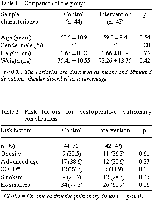

Fig. 1 - Cardiopulmonary bypass time
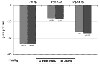
Fig. 2 - Alterations in the maximum inspiratory pressure (Ipmax) in the preoperative and on the 1st and 6th postoperative days; Pi = maximum inspiratory pressure; *p<0.05.

Fig. 3 - Forced vital capacity (FVC) and forced expiratory volume in one second (FEV1) in the preoperative period and on the 1st and 6th postoperative days *p<0.05. The variables are described as means and Standard deviations
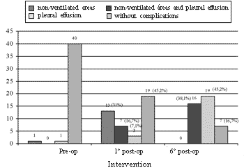
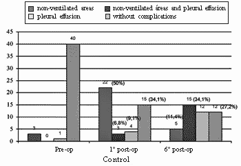
Fig. 4 - Pulmonary complications in the preoperative period and on the 1st and 6th postoperative days

Fig. 5 - Time of hospitalization
COMMENTS
In the present study, a reduction in the hospital stay can be observed among the patients of the Intervention Group, who had a mean difference of three days, when compared with the control group (p<0.05). A reduction in the hospital stay was also observed by Stein & Cassara [13] who reported a reduction in the hospital stay of patients who received physiotherapy in the preoperative and postoperative periods, when compared with the patients who did not perform physiotherapy (0.05). Semanoff et al. [14] also reported a reduction in the hospital stay for those patients who received two or more sessions of physiotherapy (ventilatory exercises, cough and precocious mobilization, as well as information about postoperative procedures) in the preoperative period of cardiac surgery. They observed that patients submitted to valve surgery who received information in the preoperative period were discharged eight days before those who were not treated with respiratory physiotherapy in the preoperative period.
Additionally, Celli et al. [15] reported a reduction in the hospital stay in the group who received guidance on ventilatory exercises (9.6 ± 3.2 days in hospital) in relation to a Control Group (13 ± 5 days in hospital). In a study performed by Healy [16] with 321 patients, in which 181 received instructions (ventilatory exercises with deep inspiration, coughing, specific explanations about the surgery in the preoperative and postoperative periods) and 140 patients who did not received any kind of intervention, the hospital stay was reduced by three to four days for the group of instructed patients.
Pulmonary function alterations occur in all patients after hours of surgical processes [17]. The reduction of the pulmonary volumes can be observed in both the Intervention and Control Groups especially from the preoperative period to the 1st postoperative day, with an increase, but without returning to the preoperative levels by the 6th postoperative day.
According to Meyers et al. [17], the pulmonary volumes (FEV1, FVC) reduced in the postoperative period [18] with a maximum decrease on the 1st postoperative day, returning to close to the preoperative levels by the 5th postoperative day [17]. The same occurred with the inspiratory muscle strength. This decreased from the preoperative period to the 1st postoperative day but there was an improvement, but without total recovery to the preoperative values by the 6th postoperative day in the Intervention and Control Groups. These tests depend on the understanding of the exercises to be performed and on the desire of the patient to collaborate in making an effort to perform the movements [19].
Thus, it is accepted that factors, such as, pain [20], alterations in the ventilatory mechanics due to the sternotomy [18] and the deleterious effects of the general anesthesia on the pulmonary function contributed to these findings. In regards to the muscle strength, sternotomy harms the stability of the thoracic wall and there is a reduction in the sanguineous support to the intercostal muscle due to the removal of the internal mammary artery, which may decrease the inspiratory muscle strength [18].
The incidence of pulmonary complications is difficult to determine from the literature, as many researchers separate these complications from the clinical significance and even the radiological significance of the disease. In additional, they depend on the type of surgery and on the individual perception of the observer about what constitutes a complication [5]. In the present study, the incidence of pulmonary radiological complications was investigated, without the clinical aspects being analysed. The presence of non-ventilated areas (atelectasis or consolidation) and pleural effusions were considered to be radiological complications. The term non-ventilated area is a concept utilized by the radiology service for those patients who present with atelectasis or consolidation in the chest radiograph.
Twenty-three (54.8%) patients of the Intervention Group and 29 (65.9%) of the Control Group developed some kind of pulmonary radiology alteration on the 1st postoperative day. Additionally, it was seen that patients of the Control Group had a greater incidence of pulmonary radiological complications on the 1st postoperative day, but without statistical significance between the two groups. On the 6th postoperative day however, there was almost no difference between the groups: 35 patients, in the Intervention Group and 32 in the Control Group developed some type of radiological alteration. The incidence of non-ventilated areas can be observed as being predominant in the Control Group in the preoperative period as well as on the 1st and 6th postoperative days.
Harm to the pulmonary function in the postoperative period is inevitable, but, sometimes, it is not clinically significant. This would justify the hospital release of many patients with radiological alterations.
According to Wilcox et al. [21], abnormalities can be analysed by chest radiograms in many patients in the postoperative period of cardiac surgery, without resolutions when these patients are discharged from hospital. The origin of pulmonary complications in the postoperative period is multifactorial and include changes in the ventilatory mechanics due to sternotomy, pain [20], hypoventilation, anesthetic effects [6], as well as retraction of the lower left lobe, postoperative gastric distension and paresis of the left diaphragmatic hemi-cupula caused by the surgery or hypothermic damage to the phrenic nerve in patients who underwent coronary artery bypass surgery with mammary anastomoses [22].
In the current study, a high incidence of pleural effusions was observed in the sample on the 6th postoperative day (72%), but with no statistical difference between the groups. From the 86 patients included in the study, 73 (84.9%) underwent coronary artery bypass grafting using the internal mammary artery (with or without the association of a saphenous vein graft). From these, 35 (83.3%) patients of the Intervention Group and 27 (61.4%), of the Control Group suffered pleural effusions by the 6th postoperative day. In a study performed by Vargas et al. [22] a high incidence of pleural effusions was also observed in patients who underwent coronary artery bypass grafting with mammary anastomosis. Thus, the high incidence of postoperative pleural effusions may be explained by the great number of patients who underwent coronary artery bypass grafting with mammary anastomosis.
Limitations of the present study
1) Radiological analysis: as was demonstrated by some authors [23-25], it is difficult to obtain precise diagnosis of pulmonary complications on chest radiographs. In a work performed by Bloomfield et al. [23], the intra-observer and inter-observer differences in respect to the description of the radiological abnormalities were clearly observed, suggesting that, when chest radiographs are analysed, the alterations should be described and diagnoses should not be made: or differential diagnoses should be suggested. In the cited study, atelectasis and consolidation were not distinctly described and presence of atelectasis or consolidation was considered as non-ventilated areas, according to the criteria utilized by the radiology service.
2) Sample calculation: when the calculation of the sample was performed for this study (90 patients) the incidence of pulmonary radiological complications was underestimated at 20% in the postoperative period. It is very clear that there were a greater number of cases and consequently a greater sample size was necessary. Particularly as patients who are submitted to coronary artery bypass grafting have many correlation factors and present with many variables, which demand a greater number of patients to obtain significant data.
Works demonstrate limitations in the methodological description, which makes it difficult to safely conclude the reliability of the findings [26]. Another aspect to be considered is the absence of uniform criteria to establish the presence of pulmonary complications and to describe the preoperative and postoperative therapeutic regimen, as well as the relative importance given to the risk factors of pulmonary complications, which may contribute to discrepancies in the results [27].
The demonstration and explanation of the importance of ventilatory exercises and early mobilization by the physiotherapist are important [16]. However, the reported postoperative complications did not have clinical relevance to the point that the patient required specific therapeutic intervention or more time of hospitalization. Thus, the greater attention given to the patient in the preoperative period can influence the faster postoperative recovery.
Based on this study, it is possible to predict that patients instructed in the preoperative period will be better prepared to collaborate with the necessities of the postoperative therapy. Their understanding of the aims of preoperative and postoperative physiotherapy, the resulting limitations of the surgical process and the proposed physiotherapeutic technique, will possibly help in the recovery and so reduce the time of hospital stay.
Thus, it is important to think of the cost-effectiveness of a preoperative physiotherapy program, of at least two sessions, which may, not only reduce the hospital stay, but reduce the hospital costs. The incidence of pulmonary radiological complications already suggests that a new sample size should be studied in order to verify the effectiveness of preoperative physiotherapy in the incidence of pulmonary radiological complications, as the size was not sufficient to demonstrate the efficacy of the treatment in the present study. On the other hand, differences were not found in respect to the maximum inspiration muscle strength (maximum PI) and forced expiratory volume in one second (FEV1), in the patients who participated in this study.
BIBLIOGRAPHIC REFERENCES
1. Verri J, Barbosa VG, Kalil PSA. Pré e pós-operatório de cirurgias cardíacas. In: Menna Barreto SS, Vieira SRR, Pinheiro CTS, editores. Rotinas em terapia intensiva. 3ª ed. Porto Alegre:Artmed;2001. p.427-34.
2. Akdur H, Polat MG, Yigit Z, Arabaci U, Ozyilmaz S, Gürses HN. Effects of long intubation period on respiratory function following open heart surgery. Jpn Heart J. 2002;43(5):523-30.
3. Hess DR. The evidence for secretion clearance techniques. Respir Care. 2001;46(11):1276-93.
4. Iglezias JCR, Lourenção Jr. A, Dallan LAO, Puig LB, Oliveira SA. Revascularização do miocárdio no paciente idoso: com ou sem circulação extracorpórea? Rev Bras Cir Cardiovasc. 2003;18(4):321-5.
5. O'Donohue Jr. WJ. Postoperative pulmonary complications. When are preventive and therapeutic measures necessary? Postgrad Med. 1992;91(3):167-70, 173-5.
6. Stiller KR, Munday RM. Chest physiotherapy for the surgical patient. Br J Surg. 1992;79(8):745-9.
7. Macedo Nedo AV, Moreschii AH. Pré e pós-operatório de cirurgia torácica. In: Menna-Barreto SS, Viera SRR, Pinheiro CTS, editores. Rotinas em terapia intensiva. 3ª ed. Porto Alegre:Artmed;2001:450-9.
8. Cuello AF, Masciantonio L, Cuello GA. Entrenamiento muscular com patrones musculares respiratórios em diferentes patologias y distribución regional de ventilación. Med Intensive. 1988;5:68-77.
9. Sciaky AJ. Educação do paciente. In: Frownfelter D, Dean E. Fisioterapia cardiopulmonar: princípios e prática. 3ª ed. Rio de Janeiro:Revinter;2004. p.355-63.
10. Sociedade Brasileira de Pneumologia e Tisiologia. I Consenso Brasileiro de Espirometria. J Pneumol. 1996;22(3):105-64.
11. Barnes TA. Core textbook of respiratory care practice. 2ª ed. St. Louis, Missouri:Mosby;1994.
12. Souza RB. Pressões respiratórias estáticas máximas. J Pneumol. 2002;28(3):155-64.
13. Stein M, Cassara EL. Preoperative pulmonary evaluation and therapy for surgery patients. JAMA. 1970;221(5):787-90.
14. Semanoff T, Kleinfeld P, Castle P. Chest physycal therapy as a preventive modality in cardiac surgery patients. Arch Phys Med Rehabil. 1981;62:506.
15. Celli BR, Rodriguez KS, Snider GL. A controlled trial of intermittent positive pressure breathing, incentive spirometry, and deep breathing exercises in preventing pulmonary complications after abdominal surgery. Am Rev Respir Dis. 1984;130(1):12-5.
16. Healy KM. Does preoperative instruction make a difference? Am J Nurs. 1968;68(1):62-7.
17. Meyers JR, Lembeck L, O'Kane H, Baue AE. Changes in functional residual capacity of the lung after operation. Arch Surg. 1975;110(5):576-83.
18. Berrizbeitia LD, Tessler S, Jacobowitz IJ, Kaplan P, Budzilowicz L, Cunningham JN. Effect of sternotomy and coronary bypass surgery on postoperative pulmonary mechanics: comparison of internal mammary and saphenous vein bypass grafts. Chest. 1989;96(4):873-6.
19. Syabbalo N. Assessment of respiratory muscle function and strength. Postgrad Med J. 1998;74(870):208-15.
20. Guizilini S, Gomes WJ, Faresin SM, Carvalho ACC, Jaramillo JI, Alves FA et al. Efeitos do local de inserção do dreno pleural na função pulmonar no pós-operatório de cirurgia de revascularização do miocárdio. Rev Bras Cir Cardiovasc. 2004;19(1):47-54.
21. Wilcox P, Baile EM, Hards J, Muller NL, Dunn L, Pardy RL et al. Phrenic nerve function and its relationship to atelectasis after coronary artery bypass surgery. Chest. 1988;93(4):693-8.
22. Vargas FS, Cukier A, Terra-Filho M, Hueb W, Teixeira LR, Light RW. Influence of atelectasis on pulmonary function after coronary artery bypass grafting. Chest. 1993;104(2):434-7.
23. Bloomfield FH, Teele RL, Voss M, Knight DB, Harding JE. Inter and intra-observer variability in the assessment of atelectasis and consolidation in neonatal chest radiographs. Pediatr Radiol. 1999;29(6):459-62.
24. Albaum MN, Hill LC, Murphy M, Li YH, Fuhrman CR, Britton CA et al. Interobserver reliability of the chest radioghaph in community-acquired pneumonia. PORT Investigators. Chest. 1996;110(2):343-50.
25. Melbye H, Dale K. Interobserver variability in the radiographic diagnosis of adult outpatient pneumonia. Acta Radiol. 1992;33(1):79-81.
26. Pasquina P, Tramér MR, Walder B. Prophylactic respiratory physiotherapy after cardiac surgery: systematic review. BMJ. 2003;327(7428):1379.
27. Thomas JA, Mclntosh JM. Are incentive spirometry, intermittent positive pressure breathing, and deep breathing exercises effective in the prevention of postoperative pulmonary complications after upper abdominal surgery? A systematic overview and meta-analysis. Phys Ther. 1994;74 (1):3-16.
 All scientific articles published at rbccv.org.br are licensed under a Creative Commons license


 Read in Portuguese
 Read in English
 Portuguese PDF
 Print
Print
 Send this article by email
 How to cite this article
 Submit a comment
 Mendeley
 Pocket