![]()
![]()
Domingo Marcolino Braile; Moacir Fernandes de Godoy
DOI: 10.5935/1678-9741.20120019
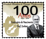
Revised edition celebrating 100 years of the birth of Professor Euryclides de Jesus Zerbini
The following article was written for "The Paths of Cardiology", of the Brazilian Archives of Cardiology in 1996. The space was coordinated by Prof. Luiz V. Décourt, whose commentary is reprinted at the end
History, to deserve that name, requires anyone who wants to develop it, not only general culture as well as broad and based special knowledge that pass through Heuristics, Bibliography, Critical and Historical Methodology and other auxiliary sciences, such as Palaeography, Diplomatic Chronology, Synthesis and Historical Exhibition [2].
We do not claim, of course, to have each of the ideal qualities required for this mister, but direct contact we had with the subject in the last quarter century and the relatively short time of existence of cardiac surgery in current use, minimizes our disability and facilitates this presentation.
It is known that in Brazil until the late nineteenth century, surgical procedures were not performed, but those simpler, which were charged to the "Barber", "barber-bleeder" or "barber-surgeon', who practiced bloodletting and scarification, applied suckers, leeches and enemas, lanced abscesses, performed curative, excised foreskins, treated the snake bites, pulled teeth, etc. The majority was composed of laymen, ignorant and humble social class [3]. Even in Europe, surgery was, in general, incipient then and in terms of cardiac approach, totally lacking. In 1882, Theodor Billroth commented that performing pericardiectomy tantamount to an act of prostitution in surgery or surgical frivolous, stating the following year that every surgeon who tried to stitch up a wounded heart should lose the respect of his colleagues. Soon, however, Ludwig Rehn, in 1896, successfully performed a right ventricular wound suture4. Even with the approach of the heart, it was at least curious an observation by Sherman, in 1902, in The American Journal of Medical Association [4], when he commented that the distance to achieve that body is no bigger than an inch, but it took 2,400 years that the surgery could go this route.
In fact, there was only little more than four decades that cardiac surgery, along the lines as we know it today began to take shape, and since then, the progress has been staggering. The scientific advances of the twentieth century have demystified the heart as seat of the soul, putting it in a hierarchical level not far from the other organs of the body. Thus began the history of cardiac surgery!
The term history comes from the Greek historía with original meaning of search, investigation or resarch [5]. In this sense will be given the primary focus of this study, or that is, the systematic search of available literature on the subject, attempting also to present it in the form of topics for related issues and not just following the chronological order, thus facilitating the perception of evolutionary aspect of the matter.
We will examine the facts relating to the open heart surgeries and those under direct vision or open. At the end will be performed some specific comments on the cardiac surgery in Brazil.
Open heart surgeries
Among the open heart surgeries, we will highlight those related to congenital heart disease, the mitral or aortic stenosis and the type of coronary artery failure.
Congenital heart disease surgery - Among the congenital heart diseases likely to total or palliative correction, since the early days of cardiac surgery, we highlight the persistence of the ductus arteriosus, tetralogy of Fallot, aortic coarctation and pulmonary valve stenosis.
Known since ancient times (it was discovered and described by Galen in the second century), the ductus arteriosus or arterial conduct only began to receive more clinical attention in the early twentieth century. Until then, several studies have been published but in general with concern purely anatomical. Munro, on 06.05.1907, based on studies posmortem, described and proposed at the Academy of Medicine, Philadelphia, ligation of the arterial conduit6. The first known attempt to perform an operation was done in London by O'Shaughnessy, and is cited by Góbich in 1945 [6]. The procedure was never implemented because it was a misdiagnosis. In fact the patient had a pulmonary artery stenosis and patent ductus arteriosus was transformed into the ligament.
The 2nd intervention with the same aim was published by Graybiel et al, in Boston in 1938 (apud Góbich, 1945) [6]. It was a complicated case with bacterial endocarditis, where there were many technical difficulties of dissection of the channel, being performed only a series of plicatures that did not produce complete occlusion and the patient died four days later. Finally, the first correction of patent ductus arteriosus was successfully performed on 08/26/1938, and was performed by Dr. Robert E. Gross, the chief resident at the time, with 33 years of age. The patient was a girl of seven years old, the canal was 7 mm in diameter and, according to Gross's report, it was closed with simple ligation of twisted silk thread, 8. It is worth noting that the first surgery was performed deliberately, without the knowledge and the absence of the chief surgeon at Brigham and Boston Children's Hospital, Dr. William Ladd, who once believed that Dr. Gross would never get permission to top realization of what he called an "extravagant adventure"[7]. Since then, the surgical correction of persistent ductus arteriosus became commonplace fact and has already been performed thousands of times worldwide, including in newborns with very low rates of morbidity and mortality.
The palliative surgery of tetralogy of Fallot, by creating an anastomosis between the subclavian artery and pulmonary artery, was due solely to the tenacity of Dr. Helen Taussig. She had observed clinical deterioration of the children with cyanotic heart disease that, once there was the spontaneous closure of the ductus arteriosus, and it warned of the possibility of surgically creating a systemic-pulmonary communications. She traveled to Boston to seek the help of Dr. Robert Gross, but the latter said that "he closed ducts and did not build new ones" [8]. Only with the arrival of Dr. Alfred Blalock at Johns Hopkins and the fact that surgeons already have past experience with anastomosis between the subclavian and pulmonary artery in an attempt of experimental production of pulmonary hypertension, it was possible the palliative approach of tetralogy of Fallot.
The 1st surgery was performed on 11.29.1944 in a girl of 15 months of age, with only 10 pounds of weight and bouts of hypoxia. The postoperative course was stormy and the child died after six months. The 2nd operation was only performed on 03.02.1945 in a girl of 11 years of age and progressed well. Until 1949, 1,000 of these surgeries have been performed at John Hopkins! [8].
With regard to aortic coarctation, McNamara and Rosenberg [9] performed an excellent historical review from which we extract some data of inaccessible literature: "In 1761, Morgagni described a case where the aorta was seen as a surprising narrowing near the heart, but this description could easily fit both the supravalvular aortic stenosis as the diffuse aortic hypoplasia or coarctation. In 1791, Paris reported the first case an unequivocal classic coarctation of the isthmus of the thoracic aorta. Le Grand, in 1835, described the 1st case report of aortic obstruction, confirmed by autopsy. On 19/10/1944, Craaford & Nylin, Sweden, performed surgery of the first case and a few months later, Gross and Hufnagel, in the United States, performed the 2nd". Although the date of the operation performed by Craaford & Nylin (October 1944) was prior to the operation performed by Gross (June 1945), the study of the latter was actually the culmination of many years of careful experimental research in laboratory animals, which demonstrated the feasibility of end-to-end anastomosis of the aorta after cross resection. Since then, a variety of techniques have been reported, including national contributions [10].
The possible correction of aortic coarctation by balloon angioplasty, reduced the number of patients referred for surgery, but the possibility of re-coarctation, particularly in neonates, makes surgical techniques are still part of the therapeutic approach of that condition. Thus, with pulmonary stenosis, the dilatation using balloon catheter has provided excellent immediate and long-term results, without significant evidence of restenosis. Thus, surgery, nowadays, can be considered to be the exception and reserved only for cases of pulmonary valve dysplasia. Historically it's worth mentioning, however, the study by Sellors (1948) [11] and Brock (1948) [12] reported that the early results with surgical success in patients with pulmonary valve stenosis and normal aortic arch, using the valvulotome by transventricular via.
Surgery of valve disease -Among the surgical treatment of valvular heart disease that deserve put attention, historically, one should mention aortic valve stenosis and mitral stenosis, given the large number of patients undergone surgery and the large number of researchers involved with the matter.
Aortic valve stenosis - The first clinical trial that is known to relieve the aortic obstruction was performed in 1913 in France, by Tuffier (cited by Margutti et al, 1955) [13], when digitally dilated the valve through the aortic wall invagination.
In 1950, Brock, issued an attempt to dilate the stenosis through an instrument inserted through the right subclavian artery previously distally sectioned and connected, after leaving through the process because of technical difficulties found [13].
In the same year, Redondo-Ramirez and Brock described independently the approach by mitral stenosis, by introduction of a finger through the left atrium, this technique has come to be known as Ramirez maneuver, being, however, abandoned due to the dangers of rupture of the septal leaflet of the mitral valve with mitral severe regurgitation [13].
The experimental study performed by Horace Smith, published in 1947, in order to cooperate with the surgical treatment of the evil that afflicted himself, not only attracted attention to the problem, as elucidated several facts. This author performed in a large series of normal dogs, valvulotomy and partial valvulectomy of the aortic valve through the aortic wall or by transventricular via, demonstrating the poor tolerance to both acute aortic insufficiency that was produced with respect to the ventricular aggression [13].
Bailey et al., in 1950 [14], impressed with the early death of Smith and the success of mitral commissurotomy, and after extensive experimental and clinical studies, concluded that the separation of the commissures by instrumental dilatation through low transventricular via seemed, on occasion, be the best process, employing it routinely.
The 1st dilatation of aortic stenosis in Brazil, with the help of Bailey dilator, was performed on 16.07.1953, at Hospital São Paulo, Paulista School of Medicine by Dr. Ruy Margutti [13].
Mitral valve stenosis - Surgical correction of rheumatic mitral stenosis dates back to 20/5/1923 when Elliot Carr Cutler and Samuel Levine, using a tenotome, successfully performed the mitral commissurotomy by transventricular via in a patient 12 years age at the Peter Bent Brigham Hospital of Harvard Medical School. The patient arrived the recovery room 1 hour and 15 minutes after starting the operation, and was discharged 12 days postoperatively. Survived by four and a half years, and died as a result of pneumococcal pneumonia. After this first success, they performed seven other operations, with new models of valvulotome, to create a "controlled" mitral insufficiency, but the results were not good, making the procedure was abandoned in 1929. The historical curiosity of the event is that the valvulectomy of the 1st patient of Cutler and Levine, was performed using an adapted tenotome, once it dealt with an emergency situation and the device was specifically developed, and was still not finished at the time of operation. In 1925, Henry Souttar at the London Hospital, performed the approach of the mitral valve through the left atrial appendage, performing the commissurotomy with the aid of his own finger. Despite the success, no other case was operated by him for lack of referral from British cardiologists at that time. The development of mitral stenosis surgery was only resumed in the mid-40s when doctors Dwight Harken and Charles Bailey, independently, started to practice valvuloplasty on a large escale [15].
Surgery for coronary heart disease - coronary heart disease surgery is perhaps one of the most experienced changes over the years. Several techniques not dependent on cardiopulmonary bypass have been proposed and used, though often with uncertain and doubtful. We collected from the Proceedings of the Ninth International Congress of the International College of Surgeons held in São Paulo-SP (Brazil), between 26/ 4 and 2/5/1954, a summary of the presentation by Dr. Charles P. Bailey [16], which well demonstrates the incipient state of surgical treatment of coronary heart disease at the time: "many methods have been suggested for the relief of angina pectoris, but with the exception of coronary artery bypass surgeries, they have been essentially palliative. The bypass operations are of three types: 1) surface revascularization, as performed by Thompson; 2) the mammary implant in the myocardium as performed by Vineberg; 3) retrograde movement of arterial blood through the coronary sinus as performed by Beck et al. We perform some Beck operations, many of which are modified according to the Kralik method. It is our opinion that this is the most effective of revascularization procedures and has both anatomical and physiological evidence confirming this impression. The clinical results after intervention with this method have been very rewarding. (...)"
The knowledge we have today about coronary heart disease, explains very well that little could be done at the time in terms of surgical procedures, since not even the appropriate diagnostic methods had yet been developed. Only with the advent of coronary angiography in the early 60s, it was possible to know with more detail the pathophysiology of the process, then starting the techniques of revascularization with cardiopulmonary bypass.
Open heart surgery
The open-heart surgery can be considered as one of the most important medical advances of the twentieth century. To get an idea of the extent of its use, one should mention that in 1994 about 2,000 surgeries were performed a day in the world, without much difficulty and with low risk, even in the age groups with a higher possibility of complications, both newborns and octogenarians. It is undeniable that this fact is of utmost importance especially when considering that the 1st open heart surgery successfully performed only happened in 09/02/1952, when Dr. F. John Lewis corrected an atrial septal defect of 2 cm in diameter, under direct vision, with interruption of flow in the cavae and moderate body hypothermia (26oC) in a girl of 5 years of age at the Hospital of the University of Minnesota (USA) [17].
Moreover, the University of Minnesota can be considered as the cradle of cardiac surgery worldwide, as there really was that great things happened. It was there also that the pioneers of cardiac surgery in Brazil began under the guidance of Dr. W. Lillehei, especially Drs Euryclides de Jesus Zerbini, Delmont Bittencourt, André Esteves Lima, Hugo Felipozzi and Domingos Junqueira de Moraes, who spread knowledge here, formed schools and made heart surgery a marker of the viability of our country.
Returning to the University of Minnesota, bold techniques developed there around 1955, became that school, in a few months, the Mecca of cardiac surgeons eager to learn and patients with hope of being healed. Words such as hypothermia, cross circulation and bubble oxygenator have become common in surgery throughout the world.
Wilson [18] provides an excellent overview of how the events unfolded at the time. It seems that it all started when Dr. Owen Wangensteen, on 01/09/1939, ligated the 1st ductus arteriosus at the University, using the technique developed by Dr. Robert E. Gross, a year earlier, at Children's Hospital, Boston. Another intensely involved in cardiac surgery in Minneapolis was Dr. Morse J. Shapiro who, being clinical, became interested in valvular diseases and created a pavilion of 40 beds for treatment of children with rheumatic fever at Lymanhurst Center for Tuberculous Children, a hospital attached to the University. Examining children with rheumatic fever, also found a large number of them with congenital heart disease. Although at first did not believe much in cardiac surgery, soon he was convinced that it was necessary to operate them before it was too late. The diagnosis of patients, especially children with heart disease, increased alarmingly and the number of beds available was too small.
In 1944 there was a club in Minneapolis that brought together the owners of movie theaters in the area called the Variety Club, which became interested in the work developed by Dr. Shapiro and decided in January 1945, raising funds of approximately $ 150,000 for construction of a Heart Hospital on the campus of the University, the first at that time, and, moreover, provide a minimum of $ 25,000 annually for its operation. At that time, movie was the main entertainment of the population, since the television had not yet emerged. The members of the Variety Club, who were in direct connection with the film producers decided to ask the Warner Brothers Studio a short film to call attention to the urgent need for a Heart Hospital in Minneapolis. The artist who urged the public to assist in the campaign was none other than Ronald Reagan, later elected President of the United States. The film was presented to thousands of viewers in hundreds of theaters throughout the northwest of the nation.
The campaign was helped by the success of the Blalock-Taussig operation, since the Heart Hospital would not only diagnose and treat children, from the medical or surgical standpoint of. Thus, it was possible to raise funds of about $ 500,000, plus the contribution from the federal government over $ 600,000 and the University with other $ 400,000. The hospital was completed in March 1951 at a cost of more than 1.5 million dollars. It had four floors overlooking the Mississippi River, linked to University Hospital by a bridge on the rooftop.
Already in 1945, when the Heart Hospital was being planned, Dr. O. Wangensteen, chief of surgery, and Dr. M. Visscher, head of the Department of Physiology, encouraged Dr. Clarence Dennis, who was Associate Professor of Surgery, to develop the artificial heartung machine that would allow open heart surgeries. With the end of the war and the return of many young people for graduation, the University of Minnesota was in a privileged position in all sectors.
In the field of surgery, Dr. Wangensteen had created a peculiar way to the training of surgeons. Each member of the department had to develop some research line and the residents had to assist them in this task. Dr. Wangensteen believed that surgeons had to learn to operate and perform other routines, but also needed to read, think and search. They could not just be intellectual parasites using ideas and methods developed by others. In fact, residents should contribute to the "heritage" of the institution.
In such an atmosphere in which physiology, surgery and research were joined, was that Dr. Clarence Dennis, in 1945, began his work with the heart-lung machine. The concept of heart-lung machine was not new. In 1931, Dr. John Gibbon, working with Dr. Edward D. Churchill, seeing a patient dying on the operating table when attempting to remove a massive pulmonary embolus imagined that if it were possible to maintain circulation and oxygenation, the patient could have been saved. His work continued at the Massachusetts General Hospital in 1934 and already in 1937, had developed a machine capable of maintaining breathing and circulation in small animals for 30 or 40 minutes. In the same year, at Jefferson Medical College in Philadelphia, Dr. Gibbon built a new machine using for the first time roller pumps, which had been introduced by Dr. Michael De Bakey in 1934. This machine enough to keep cats in circulation, but it was too small for dogs and much less suitable for humans. The advent of World War II interrupted his work.
Returning from the war, Dr. Gibbon was appointed professor at Jefferson Medical College and start to develop a more efficient extracorporeal circulation machine. In 1946, he sought help from engineers and finally one of them, IBM has built a new machine, very sophisticated with temperature, level and flow controls. Thus, in 1947, Dr. Gibbon could operate some dogs. Initially, the mortality was 80%, especially by air embolism; after years of persistent attempts, it was possible to reduce mortality to 10%.
At the same time, Dr. Dennis also tried to build an oxygenator at the University of Minnesota, studying what had been performed in other schools. The blood was passed into the cellulose tubing (used for bag sausage) in order to oxygenate it in an atmosphere of oxygen. Oxygenation was very poor and the project was abandoned. When oxygen was injected directly into the blood the oxygenation was good, but formed an enormous amount of foam. He started to use rotating vertical cylinders similar to those developed by Dr. Gibbon.
This could oxygenate blood without causing too many bubbles, so that it was possible to operate animals. Many drawings of cylinders, funnels and their association were tested, always trying to improve oxygenation and reduce blood trauma. Finally a complicated apparatus was developed, of difficult operation, cleaning and sterilization. Worse, from 64 dogs operated only nine survived. While demonstrating the reduction of hemolysis, other drastic changes continued occurring in the blood of dogs in experiments such as loss of plasma and the reduction of leukocytes and platelets, followed by intestinal hemorrhage and death of the animal. Later, another researcher, Dr. Russel M. Nelson showed that these changes resulted from contamination of equipment by bacteria.
During the following year, the oxygenator was changed again, simplifying it using screen disks, revolving slowly and over which were thrown jets of blood, according to a model described by Dr. Viking Bjork. This oxygenator was sufficient to maintain the circulation and oxygenation of a human being. The experimental work continued, always with very high mortality rate of dogs. Still, on 04/05/1951, Dr. Dennis and his colleagues used the equipment in a girl of six years old, who had a large septal defect. What impressed him most was the large amount of blood continued to flow in the heart by "Tebesius system" (sic), which forced them to inhale it and return it to the oxygenator, creating then the method used today. The child died soon after the operation, but the oxygenator performance was very good.
The surgical team consisted of 16 people (two anesthetists, four surgeons, four operators of the heartlung machine, one responsible for the blood samples, two technicians and two nurses). This was the 1st patient operated using cardiopulmonary bypass in the world, unfortunately without success. Dennis, however, was not the only experiencing difficulty. In Philadelphia, Gibbon, using the oxygenator developed by IBM and made up of fixed screens on which a layer of blood passed in an atmosphere of oxygen, still presented high mortality in experimental animals. Of the 21 dogs operated for periods of 20-90 min, 14 died. However, in 1951, an important finding emerged from the presentations of teams in Philadelphia and Minnesota at the congress of the American Surgical Association; oxygenators, now in use were sufficient to maintain oxygenation of dogs and even humans, although many animals continue dying of unknown causes.
A year earlier, in 1950, a new fact emerged from an experimental work of a Toronto's team (Canada), led by Dr. W. G. Bigelow. They showed that by lowering the temperature of an anesthetized dog at 20ºC, its oxygen consumption fell to 15% of normal. This allowed to isolate and stop the heart for about 15 min to correct intracardiac defects. As many hearts fibrillated, they developed defibrillators and pacemakers to keep the same pace. Even so, mortality of animals was too high, with undetermined causes.
In July 1951, Dr. Dennis became Professor of Surgery at the University of New York Downstate Medical Center, taking with himself the heart lung machie, by which the University of Minnesota received $ 16,000, a fund employee in further research. Dr. Dennis never operated a patient's heart. Luckily, this time, Dr. C. Walton Lillehei, appointed Associate Professor of Surgery at the University of Minnesota, was full of enthusiasm for cardiac surgery.
In 1946, Dr. Lillehei, returning from the war, made residency with Dr. Wangensteen and, in the end, was operated for lymphosarcoma, which forced wide cervicalthoracic evacuation; this, however, did not discourage him, by contrast, increased his will to live and win. During his residency, Dr. Lillehei had spent two years in the laboratory of physiology with Dr. Visscher, which gave him solid experimental basis. Often he "operated" the hearts of patients who had died with congenital diseases, trying to correct the defects. Dr. Lillehei concluded that if we could reach the inside of the heart, the operations would be relatively simple. So when Dr. Wangensteen asked him what research he wanted to develop, did not hesitate in answering: "open heart surgery", which was quickly accepted by Dr. Wangensteen.
From what Dr. Lillehei knew in 1951, the cardiopulmonary bypass systems were not yet at the stage of clinical use, were complicated, difficult to sterilize and mortality of rats was prohibitive. He concluded that some simpler way had to be developed. Based on original work of British surgeons Dr. Anthony Andersen and Dr. Frank Watson, Dr. Lillehei developed the concept of "azygos flow", which in summary is the fact that they clamped the two cavae, the azygos vein flow , which is about 1/10 of the systemic blood flow, it is sufficient to maintain the brain and other organs for about 40 min. With this, he developed a simple extracorporeal circuit, which used a lobe of the lung to oxygenate blood flow similar to the azygos vein, allowing operation of dogs without mortality. Since hypothermia lowers the oxygen consumption, he suggested that the use of hypothermia could increase the safety period.
At the same time, Dr. F. John Lewis and Mansur Taufic (Brazil) began in Minneapolis, the use of hypothermia. After much experimentation on dogs, found that irreversible ventricular fibrillation was the result of coronary air embolism. Thus, they operated 10 dogs that had previously undergone correction of atrial septal defect, closing it with "hypothermia", with the death of one dog.
In late summer of 1952, Dr. Lewis, Dr. Varco and Dr. Taufic were confident in their technique of hypothermia to the point that on 2/9/1952 operated a five year old girl, underdeveloped and suffering from an atrial septal defect. The temperature was lowered to 26oC, the chest was opened, the cava clamped for 5.5 min for the closing of communication. The child was discharged 11 days postoperatively, and this was the 1st open-heart surgery performed successfully in the world. Five minutes of cardiac arrest that would revolutionize the history of heart disease.
By the year 1953, while Dr. Lewis and Dr. Taufic were performing open heart surgery with hypothermia, Dr. Lillehei and his assistants continued their research to solve the problem of oxygenating blood during complete cardiopulmonary bypass without time limit.
In 1953, Dr. Andersen and Dr. Watson in England, published their experiments of cross circulation in dogs for periods up to 30min. The group of Dr. Lillehei, Dr. Warden and Dr. Cohen decided to develop the cross circulation with a view to clinical application, they studied the physiological variables and found that none of the "donors" in the trial died.
In March 1954, Dr Lillehei and his group felt safe enough to use the cross circulation in humans.
In the Hospital of the University of Minnesota, considered very progressive, there was strong opposition to the innovative idea of Dr. Lillehei to perform the cross circulation in humans. Dr. Wangensteen was of invaluable assistance. When a second operation was planned to be suspended last night due to the opposition, Dr. Lillehei left him a note that read: "Is our operation still standing tomorrow morning?" Dr. Wangensteen received the following response: "Dear Walt, by all means go ahead."
Thus, on 03/26/1954, the team of Dr. Lillehei performed surgery on one-year-old boy, who had spent most of his life in hospital with pneumonia and attacks of heart failure, very small, weighing only 6.9 kg and presenting often cyanotic. Catheterization showed a large ventricular septal defect. For the cross circulation, the father was chosen as a "support". The movement lasted 13min, during which Dr. Lillehei closed the VSD with a continuous suture. The normal operation developed well, as well as the postoperative period until the child developed pneumonia and bronchitis, and died 11 days after the operation. The autopsy showed marked change in the pulmonary circulation.
Without hesitation, Dr. Lillehei and his colleagues performed surgery on a 2nd patient with four years old using cross circulation on 04/20/1954, using also the father of the child as circulatory support. This patient also developed pneumonia but recovered and was discharged. In late August 1954, Dr Lillehei and his assistants had undergone eight open heart surgeries for closure of VSDs, with two deaths. Facing the seriousness of the cases, the results presented represented an unsurpassed success.
On 08/31/1954, with the experience gained in the closutr of the VSDs and the training performed in the autopsy room, Dr. Lillehei operated the 1st case of tetralogy of Fallot with total correction in a 11-year-old boy, underdeveloped and very cyanotic, having left school because of illness. During the cannulation had a cardiac arrest, but the heart started beating when cross circulation was established. The VSD was closed and relieved pulmonary stenosis. The patient was discharged two weeks later, just being able to play baseball and cycling.
On 12/03/1954, another patient with tetralogy of Fallot underwent surgery at the age of 19 years, with severe heart failure and cyanosis. The defects were corrected and the patient was discharged cured.
Until February 1955, Dr. Lillehei and his group had operated with cross circulation 32 patients with 25 survivors. None of the seven deaths resulted from cross circulation. One of the deaths were due to complete AV block.
With repetition of the cases, the cross circulation became easier with the use of a cannula in each cava and a venous reservoir. The flow was maintained 30- 40% of normal cardiac output at rest. To facilitate visualization within the heart chamber, a tourniquet was applied to the aorta, which was pressed intermittently to reduce the blood within the heart.
In April 1955, Dr Lillehei presented the results of nine operations for tetralogy of Fallot with five survivors in the Congress of the American Surgical Association in Philadelphia. The survivors had heart almost normal! During the discussion, Dr. Alfred Blalock, with all his importance, said: "I never thought to live long enough to see the day when this type of surgery could be performed, I congratulate the group of Minnesota by its imagination, courage and dedication". However, Dr. Blalock suggested that the ultimate solution to support the circulation during surgery would be the heart-lung machine developed by Dr. Gibbon and not cross circulation.
Dr. Gibbon had operated the 1st patient with cardiopulmonary bypass in early 1953, but the patient died. In May 1953, Dr. Gibbon operated two patients with atrial septal defect, with complete success, opening the patient's heart under cardiopulmonary bypass and the vast field of cardiac surgery, although it has not been given prominence at the time, perhaps because with hypothermia such operations were being performed routinely by Dr. Lewis and Dr. Taufic, Dr. Gibbon was never able to repeat his feat and, after five unsuccessful attempts, he abandoned heart surgery. Although no comment was done at the meeting in Philadelphia, Dr. Lillehei knew he had at the University of Minnesota a heart-lung more efficient, safer and much simpler than all the sophisticated machinery developed by Dr. Gibbon, Dennis and others. All these oxygenators based on the principle of forming a thin layer of blood over a large surface placed under an atmosphere of oxygen. But another way to create a large interface between the oxygen and blood could be achieved by bubbling oxygen directly into the blood.
In 1950, Dr. Leland C. Clark Jr et al, working at Antioch College in Yellow Springs (Ohio), developed a small bubble oxygenator. Other attempts showed that the method of buble oxygenation was too slow and with a great tendency to foam. Dr. Clark was able to demonstrate that it was possible to eliminate the bubbles passing the blood through a tube with sticks or glass beads treated with DC antifoan A. This was a silicone compound developed by Dow Corning Company used for frying potatoes and that is still used today in cardiopulmonary bypass.
In 1952, Dr. Clark et al had developed an oxygenator capable of keeping more than 20kg animals in extracorporeal circulation.
In 1954, Dr. Richard A. DeWall, a young doctor, was initially hired as responsible for cross circulation at the University of Minnesota, after accepted as a resident. This did not happen because of his grades were not sufficient for the requirements of the University, despite the desire of Dr. Lillehei and Wangensteen. When Dr. DeWall received the news, suggested Dr. Lillehei to hire him as coach of laboratory animals. Both Dr. Lillehei as Dr. Wangensteen accepted the idea and Dr. DeWall continued with the same previous activities, with the only difference that received a payment slightly higher than residents. As a research project, which was compulsory at the University, Dr. Lillehei suggested Dr. DeWall he could work in the bubble oxygenator. He asked him also that not to worry about the previous publications and restarted all the research, from the beginning.
Dr. Lillehei and Dr. DeWall had plastic tubes (PVC) of a firm that made the tubes for mayonnaise manufacturers. In the mayonnaise manufacture, formation of foaming was also a major problem that had been solved smearing the interior of the tubes with the DC antifoan, the same product used many years ago by Dr. Clark et al. Without knowing the work of Dr. Clark, Dr. Lillehei and Dr. DeWall, thought that silicone DC antifoam A was good for mayonnaise and should be good for the blood. Even with antifoan A, there were still some bubbles in the blood. It was when Dr. DeWall was thought to create a helical tube of PVC so that the bubbles would be "pushed" upwards, while the more dense blood without bubbles would be addressed to the bottom of the PVC spiral.
In the winter of 1954, Dr. DeWall performed surgery on about 70 dogs using various techniques and revealing details such as the need to heat the blood, which he reached to manage the PVC spiral dipping into a container of warm water. The connections of the tubes and filters have been progressively improved in such a way that, in May 1955, Dr. DeWall and Dr. Lillehei believed to have an oxygenator ready for human use.
On 05/13/1955 used the oxygenator of Dr. DeWall for the 1st time in a three year old child with VSD and pulmonary hypertension; the operation developed well, but the patient died 18 hours later.
In August 1955, they had used the oxygenator in seven children from 19 months to 7 years old, with only two deaths. All seven children woke up immediately after the operation. The two deaths were not related to cardiopulmonary bypass. Oxygenators created by Dr. DeWall have been improved with use. But the cross circulation, which was allowed to advance one step on the long walk from the heart surgery, it was finally abolished.
Unlike the complex oxygenator of Dr. Gibbon, with many moving parts, the bubble oxygenator of Dr. DeWall was elegantly simple, being constructed of plastic tubing used for the food industry, cheap and disposable, and autoclavable.
In May 1956, Dr. Lillehei et al reached the milestone of 80 operations with the bubble oxygenator. Within two years, more than 350 patients had been operated. The introduction of the bubble oxygenator permitted the expansion of cardiac surgery throughout the world, where there was a well-equipped hospital and doctors with preparation and willingness to make it a reality. In Brazil, Drs. Drs. Hugo Felipozzi, Adib Jatene, Valdir Jazbik, Domingos Junqueira de Moraes, Marcos Cunha, Hélio Magalhães, Otoni Moreira Gomes e Domingo Marcolino Braile, among others were dedicated to the development of oxygenators and extracorporeal circulation systems.
The attempted use of extracorporeal membrane oxygenation systems allowed, after innumerable mistakes and successes in modern membrane oxygenators, the approach of progressively more complex heart disease with good results in the short and long term. Tens of surgeons may also be mentioned, each with a significant contribution to the development of cardiac surgery. This enumeration would be tedious and probably difficult to hold the reader's memory.
We believe, however, that the emphasis on the name of Dr. Nina Starr Braunwald, can serve as a beacon to illuminate this vast gallery of remarkable people. Dr. Nina Braunwald was born in New York in 1928 and was the first woman to hold an open-heart surgery. Among other firsts, she was also the first woman to be elected to the American Association for Thoracic Surgery. In the late 50's she developed a flexible polyurethane mitral valve prosthesis with Teflon tendinae cordae, implanting it into dogs and, in 1960, led a team that first used this prosthesis for mitral valve replacement in humans. The patient survived the surgery and remained clinically well for several months. Dr. Braunwald also developed a ball covered mechanical prosthesis, the Braunwald-Cutter prosthesis, which came to be implanted in several patients. She was also pioneer in the use of tissue culture techniques, in order to create non-thrombogenic surface for prostheses and assisted circulation devices. Dr. Nina Starr Braunwald died on 08/05/1992 [19].
In Brazil, many devoted to the field of biological and mechanical prostheses, leading the country towards self-sufficiency and international prominence.
Heart Transplant
Both the scientific community and the general public were noticeably shaken when, on 12/03/1967, Dr. Christiaan Barnard in Cape Town, South Africa, performed what until then was considered the first heart transplant in human. Thereafter, an avalanche of other cases was being operated, so that in a compilation of Haller and Cerruti, until October 1968, they accounted more than 60 transplants around the world [20].
The 1st heart transplant in Latin America (17th in the world), was performed at Clinics Hospital in São Paulo, Brazil, by Dr. Euryclides de Jesus Zerbini on 05/26/ 1968. The receiver was a man of 32 years, cowboy and bearer of dilated cardiomyopathy, probably due to Chagas disease. That fact caused a great impact at the time, being matter of emphasis in both scientific and the lay press, with several newspapers and magazines occupy almost the whole space with the subject or opening extras (Folha de São Paulo, year 48, No. 14,225, Supplement I, 05.27.1968).
Cardiac transplantation is now a reality, with more than 25,000 cases recorded up to 1993 [21] and everything indicates that this number will keep increasing. In this respect, it may be considered prophetic words of Sir Peter B. Medawar (Nobel Prize in Medicine, 1970), on 09/11/1968, at the II International Congress of the Transplantation Society in New York [22]: "The organs transplantation will be assimilated into clinical practice ... and there is no need to philosophize about it. This will be true for the simple and sufficient reason that people are constituted in such a way that they would rather live than die". We believe that this would not be excessive considering this as a valid justification for the development of heart surgery as a whole.
Cardiac surgery in Brazil
The Brazilian participation in each of the topics addressed, has been duly pointed out. One should consider, however, a more generic approach to characterize the importance of cardiac surgery in our midst. Thus, in 2011 were performed in Brazil 100,000 heart surgeries, being 50,000 using CEC with more than half of them for CABG, with results comparable to those of international literature. The surgeries were performed in more than 170 centers distributed in all Brazilian states with the participation of more than 1000 surgeons members of the Brazilian Society of Cardiovascular Surgery.
There is no doubt that cardiac surgery in Brazil can find today at an equivalent level of large centers, with multiple poles featured throughout the national territory. It would therefore unfair to emphasize names, because many contributed so that Brazil could occupy the prominence position in the concert of nations. However, it is impossible to not mention the founders of the specialty in Brazil: Hugo Felipozzi, Euryclides de Jesus Zerbini, Domingos Junqueira de Moraes and André Esteves Lima, who, with idealism and courage, gave example for the Brazilian cardiac surgeon was forged with ethical and moral principles, which we expect will persist throughout the career of all who embrace this noble career that unites science, art and inventive power. That feeling of compassion for patients is a constant, leading back to normal life those who lost their health in the exercise of citizenship.
We will always pay homage to Prof. Zerbini!
As an example of this generation, we take the liberty to quote only a disciple to represent us, thanks to his performance as a surgeon, teacher, scientist and public figure, Full Professor Adib Domingos Jatene. Among his contributions, the anatomic correction of transposition of great vessels is a sample of his stimulating qualities for Brazilian surgeons be recognized internationally.
Following, celebrating 100 years of Birth of Professor Zerbini, the esteemed readers will find a clipping of the odyssey that represented the implementation of cardiac surgery at the Faculty of Medicine, University of São Paulo, the most important Brazilian universitiy. Its protagonist was Professor Euryclides de Jesus Zerbini, whose history is narrated in the biography written by his disciples Noedir G. Stolf and Domingo M. Braile.
REFERENCES
1. Políbios. História - Livro I, (seleção, tradução, introdução e notas de Mario da Gama Kury). Brasília: Universidade de Brasília; 1985.
2. Blanco RR. Técnica da Pesquisa Científica, vol I e II. São Paulo: Cupolo Ltda; 1978.
3. Santolho LC. História Geral da Medicina Brasileira, vol 1º. São Paulo: Humanismo Ciência e Tecnologia Hucitec Ltda e Universidade de São Paulo; 1977.
4. End A, Wolner E. The Heart: location of the human soul - site of surgical intervention. J Card Surg. 1993;8(3):398-403. [MedLine]
5. Kury MG. In: Herôdotos. História (tradução do grego, introdução e notas de Mario da Gama Kury). Brasília: Universidade de Brasília; 1985.
6. Góbich E. El conducto arterioso - estudio anatomico, fisiologico, clinico-quirurgico. Buenos Aires: Prensa Medica Argentina; 1945.
7. Clathworthy Jr HW, Robert EG. A memorial surgical rounds 1989:55-68 apud Gott VL -And it happened during our lifetime... Ann Thorac Surg. 1993;55(5):1057-64. [MedLine]
8. Gott VL. And it happened during our lifetime... Ann Thorac Surg. 1993;55(5):1057-64. [MedLine]
9. McNamara DG, Rosenberg HS. Coarctação de la Aorta. In: Hamish Watson - Cardiologia Pediatrica. Barcelona: Salvat; 1970. p.188.
10. Mendonça JT, Carvalho MR, Costa RK, Franco Filho E. Coarctation of the aorta. A new surgical technique. J Thorac Cardiovasc Surg. 1985;90(3):445-7. [MedLine]
11. Sellors TH. Surgery of pulmonary stenosis; a case in wich pulmonary valve was successfully divided. Lancet 1948;1(6513):988. [MedLine]
12. Brock RC. Pulmonary valvulotomy for the relief of congenital stenosis; report of three cases. Br Med J. 1948;1(4562):1121. [MedLine]
13. Margutti R, Borges S, Campos Filho CM, Gallucci C, Branco Jr LB. Tratamento cirúrgico da estenose valvular aórtica. Rev Paulista Med. 1955;46:82-91.
14. Bailey CP, Glover RP, O'Neil TJ, Redondo-Ramirez HP. Experiences with the experimental surgical relief of aortic stenosis J Thorac Surg. 1950;20(4):516-41. [MedLine]
15. Cohn LH. The first successful surgical treatment of mitral stenosis; The 70th anniversary of Elliot Cutler's mitral commissurotomy. Ann Thorac Surg. 1993;56(5):1187-90. [MedLine]
16. Bailey CP. Surgical treatment of coronary artery disease Anais do IX Congresso Internacional do Colégio Internacional de Cirurgiões - vol IV - Parte Científica. São Paulo, SP - Brasil; 1954.
17. Lillehei CW. The Society Lecture. European Society for Cardiovascular Surgery Meeting, Montpellier, France, September 1992. The birth of open-heart surgery: then the golden years. Cardiovasc Surg. 1994;2(3):308-17. [MedLine]
18. Wilson LG. The development of cardiac surgery at Minnesota 1940-1960. In: Wilson LG - Medical Revolution in Minnesota: a History of the University of Minnesota Medical School. St. Paul: Midewiwin Press; 1989.
19. Waldhausen JA. In Memoriam: Nina S. Braunwald, 1928-1992. Ann Thorac Surg.1993;55(5):1055-6. [MedLine]
20. Haller JD, Cerruti MM. Heart transplantation in man: compilation of cases. January 1, 1964 to October 23, 1968. Am J Cardiol. 1968;22(6):840-3. [MedLine]
21. Kaye MP. Pediatric thoracic transplantations: the world experience. J Heart Lung Transplant. 1993;12(6 pt2):S344-50. [MedLine]
22. Kantrowitz A, Haller JD. Symposium on human heart transplantation. Introduction. Am J Cardiol. 1968;22(6):761. [MedLine]
23. Braile DM. The health care system in Brazil. Curr Surg. 1991;48(6):361-4.
PATHS OF CARDIOLOGY
Coordinator - Louis V. Décourt
The article by Braile and Godoy focuses an impressive history. It is known that surgery of the chest remained in long period of absence due to peculiar conditions of local viscera. Also in 1941, the English translation of the great History of Medicine, by Castiglioni, it was highlighted the fact that, "except for the drainage of empyema and lung abscess, thoracic surgery is far behind other areas of the body".
And the situation worsened when it was considering interventions on the heart, either by technical difficulties, either by the significance of the organ as untouchable structure in the physical and spiritual constitution of the body. It must be remembered that Prof. Theodor Bilroth (1829-1894), cited in the current study as relentlessly critical of interventions on the heart, was a great surgeon, founder of School of Surgery in Vienna, and certainly one of the pioneers in the modern surgery of the abdomen. Therefore, he was peculiar interpreter of medical ideas of the end of the last century*.
Subsequently, in little more than four decades, there was an exquisite evolution fruit of dreams and also tenacity and unwavering perseverance. There was, throughout the period, the right attitude of the researchers. There were questionable adventures, unjustified improvisations, unacceptable bold but sensible attitudes based on a double orientation: the constant search for experimental bases always instructive and obedience and clinical, functional and safe rationale. And this behavior was not affected by initial failures (Bailey) and even serious illness of a researcher (Lillehei).
I followed closely this extraordinary evolution through my brotherly work of my Clinics with Professor Zerbini's team. And I have seen the transformation of the past taboo in today's high corrective routine.
In this study, teachers Braile and Godoy bring broad and thorough overview with illuminating details about phases of failures, expectations and wins until the great current position. And its reading provides us with an image of the medicine of our time, so it represents a persistent, conscientious and fruitful struggle.
Article originally published in the Brazilian Archives of Cardiology, Volume 66:(1), in 1996. Reproduction authorized by the Editors.
* Editor's Note: The author referred to the nineteenth century, since the article is from 1996.
 All scientific articles published at rbccv.org.br are licensed under a Creative Commons license


All rights reserved 2017 / © 2024 Brazilian Society of Cardiovascular Surgery
DEVELOPMENT BY ![]()
 Read in Portuguese
 Read in English
 Portuguese PDF
 Print
Print
 Send this article by email
 How to cite this article
 Submit a comment
 Mendeley
 Pocket