RESUMO
OBJETIVO: Aferir a qualidade de vida de pacientes com insuficiência cardíaca refratária, inscritos como candidatos a transplante de coração. MÉTODOS: Estudo prospectivo, descritivo, transversal de 18 pacientes, com média de idade de 52 anos, em acompanhamento ambulatorial pré-transplante, de um hospital público e vinculado ao ensino do Município de São Paulo. A qualidade de vida foi avaliada por meio do questionário genérico The Medical Outcomes Study 36-item Short-Form Health Survey (SF-36), com a finalidade de avaliar aspectos relativos a função, disfunção, desconforto físico e emocional. RESULTADOS: Dessa amostra, 14 (77,8%) pacientes eram do sexo masculino e quatro (22,2%), do sexo feminino; 14 (77,8%) dos pacientes foram classificados segundo tipo funcional IV e quatro (22,2%) em tipo funcional III (New York Heart Association); 17 (94,4%) encontravam-se em estágio D e um (5,6%) em estágio C (American Heart Association/ American College of Cardiology). As médias obtidas na avaliação das escalas do SF-36 foram: capacidade funcional 38%, dor 49%, estado geral de saúde 49%, vitalidade 39%, aspectos sociais 53%, aspectos emocionais 43% e saúde mental 54%. CONCLUSÃO: A qualidade de vida dos pacientes com insuficiência cardíaca terminal é considerada muito ruim; provavelmente pior que em muitas outras entidades mórbidas mais comuns. Os aspectos, social e mental são os menos afetados, sendo os mais comprometidos, a vitalidade e a capacidade funcional.
ABSTRACT
Objective: To assess the quality of life of patients with refractory heart failure disease as candidates for heart transplant. Methods: A transversal, descriptive and prospective study with 18 adult patients, with mean age of 52 years under pre-transplantation outpatient follow-up at educational and public hospital in São Paulo town. The quality of life was assessed by reference to "The Medical Outcomes Study 36-item Short-Form Health Survey" (SF-36) generic questionnaire in order to assess the aspects in relation to the function, dysfunction, physical and emotional uneasiness. Results: According to this group, 14 (77.8%) of these patients were male and four (22.2%) female; 14 (77.8%) of them were classified as functional class IV and four (22.2%) as functional class III (New York Heart Association); 17 (94.4%) of them were at stage D and one (5.6%) at stage C (American Heart Association/American College of Cardiology). The mean results obtained from the assessment of SF-36 scales were: functional capacity 38%, pain 49%, health general condition 49%, vitality 39%, social aspects 53%, emotional aspects 43% and mental health 54%. Conclusion: The quality of life of patients presenting terminal heart failure is considered to be very bad; it is likely to be worse than in many other more common morbid entities. Both mental and social aspects are least affected, on the other hand the vitality and functional capacity are the most affected.
INTRODUCTION
Although relatively uncommon and with an incidence around 0.4% to 5% [1], the infected sternal wound dehiscence presents significant rate of mortality, morbidity and hospital costs. Mortality can vary from 6% to 70% [2].
Different techniques have been used to treat this serious complication. Among the methods of maneuver are included resutures and continuous irrigation [3,4], two-stage treatment and dead space obliteration using muscle flaps (rotated or translated using vascular pedicles, including the major pectoralis, rectus abdominis and the large dorsal) [3 -9].
Due to the importance of this type of complication, this issue has attracted cardiovascular and plastic surgeons, for over forty years [1-3,10-20]. In our institution, since 1986, we began to treat - consecutively - the sternal wound infection (with debridement, wide drainage and immediate closure) with bilateral pectoralis major myocutaneous advancement flap, whose initial results were published in 1993 [21]. This technique has been described, in the same way, by several authors [22.23], even in children previously undergone treatment of congenital heart disease [24,25].
The aim of this study is to report our experience in this current series of 13 non-selected patients who had undergone treatment with the standard protocol initiated in 1986. This protocol aims at early diagnosis and cure of the infection and functional and aesthetic recovering, providing the patients capacity for normal activity.
METHODS
Between January 2000 and July 2007, at Clementino Fraga Filho Hospital-FM-UFRJ and Aloysio de Castro State Institute of Cardiology, RJ, among 1972 heart surgeries, 13 (0.67%) patients presented deep sternal wound infection and dehiscence. All patients with this complication were treated by the same protocol. Most patients had undergone CABG, including left internal mammary artery in all patients (11), surgery for valve replacement (one), and a child had undergone total correction of
atrioventricularis communis defect.
Twelve patients had been previously undergone single sternotomy and one patient two sternotomies. There was male predominance, with only five female patients. The age ranged between two months and 67 years. The risk factors were: chronic obstructive pulmonary disease, long-term ventilation in ventilatory assistance, long duration of cardiopulmonary bypass, diabetes mellitus, prior surgery and systemic infection.
From a clinical point of view, the arising of characteristic signals on the wound, such as redness, hyperemia, purulent collection and mostly sternal instability, were sufficient to induce the diagnosis of infection or sternal dehiscence. The CT and chest radiography were of great value in the diagnosis of deep sternal wound infection, however, the continuous and daily search of this patient's complication in the early postoperative period is crucial.
This study was submitted and approved by the Research Ethics Committee of the State Institute of Cardiology at the meeting on November 12, 2008 under number 2008/22.
Surgical technique
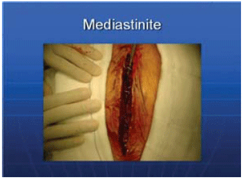
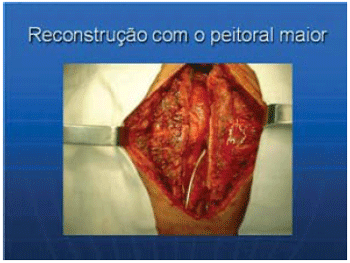
Once confirmed the diagnosis of sternal infection (Figure 1), we performed early intervention that consisted of incision and excision of 0.5 cm of each wound edge, removal of steel wires, rigorous debridement of necrotic tissue, until occurrence of tissue bleeding. Samples of mediastinal fluid and bone tissue for culture and antibiogram were collected. Careful and extensive cleaning of the wound was performed using saline solution at 37º degrees Celsius, in a procedure similar to that practiced in management of exposed wounds (Figure 2).

Fig. 1- Total dehiscence of the sternal wound after CABG.

Fig. 2 - Sternal wound after total debridement and major pectoralis bilateral dissection with anterior mediastinal drain
We began dissection and elevation of the left pectoralis major muscle and corresponding subcutaneous flap through diathermy, from the median line along the costal grid, up to two thirds of the anterior chest wall, preserving intact the humeral insertion, thoracoacromial vascular bundle and pectoralis minor muscle. At inferior plane, the pectoralis major muscle was raised - including the anterior rectus fascia - and down up to the xiphoid process. The upper segment of the anterior rectum has been raised with the pectoralis major muscle, while the abdominal rectus was not involved in the dissection. The hemostasis of intercostal perforating arterial branches was accurately performed.
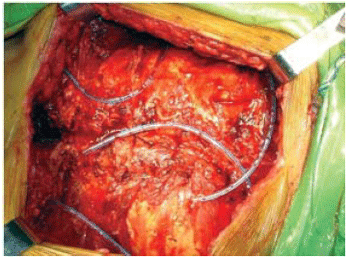
The dissection was performed - similarly to the right pectoralis major muscle. A drain tube (Number 34) was placed in the anterior mediastinum. A catheter was installed in the skin of the upper edge of the sternal wound by counter-opening for instillation of saline solution with antibiotics, followed by osteosynthesis using steel wire (number 5). In patients with fracture or asymmetry of the sternum incision, osteosynthesis was performed using the Robiscek technique. Two suction drains were placed below the muscular plane upon the costal grid for drainage of the large area of detachment of the musculoaponeurotic layer (Figure 3).

Fig. 3- Costal grid with sternal resuture and drains positioned upon the costal grid. Muscle-cutaneous flap of the major pectoralis bilaterally withdrawn
Approximation of the major pectoralis and fascia was performed, including part of the anterior rectum, using monofilament number 2 (polyglycolic acid). Sutures were performed 2 cm from the muscle-aponeurotic edge with interrupted sutures and without any tension. The skin and the major pectoralis muscle-cutaneous flap were sutured, in interrupted sutures using 00 mononylon thread. It is of great value the clinical support of associated diseases and the administration of antibiotics, as the antibiogram sensitivity.
Postoperative care
Continuous irrigation of 100 ml per hour for 24 h is performed. If at the end of 48 hours the leaving of liquid presents clear without fibrin, clot or debris, the irrigation flow is reduced to 75 or 50 ml and finished within a week. If after finishing of irrigation there is persistence of drainage, the drains will continue to be placed until all liquid had left. Patients continue to receive appropriate intravenous therapy while hospitalized and until the third week in case of need for vancomycin. As the majority of infections produced by Staphylococcus species, the antibiotic of choice was the vancomycin and up to the third week.
For irrigation of the topic solution, we used 1 g/1000 ml cefazolin or vancomycin or gentamicin as the antibiogram and sensitivity. The wound should be cleaned twice daily using chlorhexidine, and the monofilament stitch of the suture of skin and subcutaneous cell tissue should remain for three weeks.
RESULTS
The mean time of surgery was 95 minutes. The volume replacement with concentrated red blood cells was not significant. All patients were referred for intensive care unit and 12 were extubated in the first 12 or 24 hours. The hospital discharge occurred on average at the 25th postoperative day. All patients underwent surgery during the early postoperative period of the first surgery and only one patient underwent surgery after three months presenting chronic infection (type III) due to methicillin-resistant Staphylococcus. The surgery was successfully performed and the patient was discharged from hospital on the 18th postoperative day. The predominant bacteria in this series was of
Staphylococcus species (eight), followed by
Streptococcus epidermitis (two), and a case of
Serratia marcescens and two negative cases. The drains were removed only when the drainage flow reached the limit zero. There was one (7.6%) death during hospitalization, on the 16th day of the intervention on the sternal wound due to systemic infection. There was suture dehiscence after repair. Four patients required surgical reintervention to treat residual seroma or localized infection.
At the 12th day of surgical repair of the sternal wound an obese patient with diabetes mellitus presented abundant secretion, on which the
Serratia marcescens bacteria grew. The patient undergone then a second intervention, on which stability of the sternum and pectoralis major muscle was found - used in the wound repair. New debridement and drainage were performed. The patient evolved well and was discharged from hospital on the 20th day after the second intervention.
The patients continued to receive appropriate intravenous therapy. As the majority of infections produced by species of
staphylococcus, the antibiotic of choice was vancomycin, which was extended until the third week. For irrigation of the topical solution was used 1g/1000ml cefazolin or vancomycin or gentamicin as the antibiogram sensitivity.
The aesthetic and functional recovery of this procedure have been excellent. However, one patient complained of mild limitation of movements of shoulders, which stopped at the end of three months of physiotherapy.
DISCUSSION
To treat mediastinitis and sternum dehiscence, since the 80's, many surgeons began to perform transposition of the pectoralis major flaps, anterior rectus or epiplon with vascular pedicle for obliteration of dead space [3-9]. In 1993, we presented our experience started in 1986, in the treatment of infected sternotomy on which we used a new approach - single stage - using the pectoralis major muscle and the corresponding subcutaneous flap, taken bilaterally to the midline. We have used this procedure routinely and systematically [21.26]. Other reports have been published with the same technique and with good results [22-25]. Unlike the techniques of muscle flaps - translated or rotated with vascular pedicles [11-12] - the technique of advancement of the major pectoralis - bilaterally - is efficient, requires no additional incision and offers full aesthetic and functional recovery of the anterior chest. There are several classifications of sternal infection; we prefer the classification reported by Pairolero et al. [6], who subdivide the infection into three types:
Type I - occurs within the first week of the sternotomy. It is characterized by serosanguineous drainage without cellulitis and osteomyelitis. Type II - occurs between the second and fourth week of sternotomy and is always accompanied by purulent secretion, cellulitis, and often there is osteomyelitis. Type III - the infection is late and may arise over months or years after sternotomy. Such infections are chronic, with localized cellulitis, osteomyelitis and chondritis, and presence of mediastinitis is rare.
The patients in our series were of type I and II and only one patient was classified as type III, methicillin-resistant
Staphylococcus. The surgery was successfully performed this patient was discharged from hospital on the 18th postoperative day. In addition to the predominant bacteria in hospital infections such as species of
Staphylococcus, Streptococcus epidermitis, Pseudomonas aeruginosa and other, we had a case with presence of
Serratia marcescens bacteria. This bacteria has been found recently as the germs responsible for the nosocomial infection [23.27].
In our experience, early diagnosis and intervention contributed significantly to reduce the extent of tissue necrosis. This opinion is shared by other authors [20]. Most surgeons who uses this technique prefers a single stage approach of this complication [11,12, 21-23,28] and, to a lesser extent, in two stages [20]. In the last ten years, the use of the pectoralis major muscle has contributed to the high decrease in the 30-days operative mortality rate about 7.9% to 9.5% [23.26]. Or in some reports with no hospital 30-days hospital mortality [4.20].
In addition to other advantages, this procedure can be used in patients who underwent CABG using internal thoracic artery [20-22,23]. It is, however, contraindicated in case of hemodynamic instability and surgical site infection, as reported previously [21]. The risk factors in mediastinitis have been reported frequently, for which we must be careful and make the necessary prevention [17-19].
The use of mediastinum drainage under high vacuum pressure with polyurethane foam have been promising in the treatment of primary sternal infection after median sternotomy [16]. This procedure was considered effective according to a multicenter study involving 79 institutions in Germany with 90,000 patients who underwent heart surgery in 2007 on which 2000 (0.45%) cases of mediastinitis were found [29]. However, this procedure can only be used in patients with intact pleura and it is not free from complications such as pleural rupture and fall of cardiac output [15].
CONCLUSIONS
In the sternal wound infection involving deep planes or with post-sternotomy dehiscence, we recommend single stage management, using bilaterally the pectoralis major muscle and myocutaneous flap sutured to the midline. This behavior is efficient and can reduce the morbidity, restoring the aesthetics and anatomy of the anterior region of the chest.
REFERÊNCIAS
1. Rossi Neto JM. A dimensão do problema da insuficiência cardíaca do Brasil e do mundo. Rev Soc Cardiol Estado de São Paulo. 2004;14(1):1-8.
2. Douglas CR. Fisiologia do coração. In: Douglas CR, editor. Tratado de fisiologia aplicada às ciências da saúde. 1ª ed. São Paulo:Robe;1994.p.555-600.
3. Pereira WL. Qualidade de vida após o transplante cardíaco: análise de pacientes operados na Universidade Federal de São Paulo [Tese de doutorado]. São Paulo:Universidade Federal de São Paulo;2000.
4. Araújo DV, Tavares LR, Veríssimo R, Ferraz MB, Mesquita ET. Custo da insuficiência cardíaca no Sistema Único de Saúde. Arq Bras Cardiol. 2005;84(5):422-7. [MedLine]
5. Barroso E. Organização do transplante cardíaco no Brasil e no Estado do Rio de Janeiro. Rev SOCERJ. 2002;15(3):131-4.
6. Timerman A, Pereira MP. Tratamento atual da insuficiência cardíaca congestiva. Rev Soc Cardiol Estado de São Paulo. 2000;10(1):65-75.
7. Ferraz MB. Qualidade de vida: conceito e um breve histórico. Jovem Médico. 1998;4:219-22. In: Pereira WL. Qualidade de vida após o transplante cardíaco: análise de pacientes operados na Universidade Federal de São Paulo [Tese de doutorado]. São Paulo:Universidade Federal de São Paulo;2000.
8. Nobre MRC. Qualidade de vida. Arq Bras Cardiol. 1995;64:299-300. [MedLine]
9. Stolf NAG, Sadala MLA. Os significados de ter o coração transplantado: a experiência dos pacientes. Rev Bras Cir Cardiovasc. 2006;21(3):314-23. Visualizar artigo
10. Branco JNR. Transplante cardíaco: a experiência da Universidade Federal de São Paulo [Tese de Livre-Docência]. São Paulo:Universidade Federal de São Paulo;1997. In: Machado RC. Identificação e caracterização de cuidadores de candidatos a transplante do coração: análise de amostra de pacientes do ambulatório da UNIFESP [Dissertação de Mestrado]. São Paulo:Universidade Federal de São Paulo;2007.
11. Ciconelli RM, Ferraz MB, Santos W, Meinão I, Quaresma MR. Tradução para a língua portuguesa e validação do questionário genérico de avaliação de qualidade de vida SF-36 (Brasil S.F.- 36). Rev Bras Reumatol. 1999;39(3):143-50.
12. Branco JNR, Costa ISEA, Lobo Filho G, Moraes CR. I Diretrizes da Sociedade Brasileira de Cardiologia para transplante cardíaco: IX. Organizaçäo do transplante cardíaco no Brasil. Arq Bras Cardiol. 1999;73(Suppl 5):56-7. [MedLine]
13. Associação Brasileira de Transplante de Órgãos (A.B.T.O.). Disponível em: <http://DTR2001.saude.gov.br/transplantes. Acesso em: 6 nov.2007.
14. Kadri T, Marinelli I, Franken RA, Rivetti LA. Terapia e evolução do candidato a receptor do transplante cardíaco. Rev Soc Cardiol Estado de São Paulo. 1995;5(6):624-35.
15. Bayley KB, London MR, Grunkemeier GL, Lansky DJ. Measuring the success of treatment in patient terms. Med Care. 1995;33(4 Suppl):AS226-35. [MedLine]
16. DATASUS. Morbidade hospitalar do SUS. Ministério da Saúde - Sistema de Informações Hospitalares do SUS (SIH/SUS) 2007. Ref. Type: Electronic Citation.
17. Guimarães JI, Mesquita ET, Bocchi EA, Vilas-Boas F, Montera MW, Moreira MCV, et al. II Diretrizes da Sociedade Brasileira de Cardiologia para o Diagnóstico e Tratamento da Insuficiência Cardíaca. Arq Bras Cardiol. 2002;79(Supl. IV):1-30. [MedLine]
18. Rabelo ER, Aliti GB, Domingues FB, Ruschel KB, Brun AO. O que ensinar aos pacientes com insuficiência cardíaca e porquê: o papel dos enfermeiros em clínicas de insuficiência cardíaca. Rev Latinoam Enferm. 2007;15(1):165-70.
19. Machado RC. Identificação e caracterização de cuidadores de candidatos a transplante de coração: análise de amostra de pacientes do ambulatório da UNIFESP [Dissertação de Mestrado]. São Paulo:Universidade Federal de São Paulo;2007.
20. Guillemin F, Bombardier C, Beaton D. Cross-cultural adaptation of health-realted quality of life measures: literature review and proposed guidelines. J Clin Epidemiol. 1993;46(12):1417-32. [MedLine]
21. Frota Filho JD, Lucchese FA, Blacher C, Halperin C, Jawetz J, Lúcio EA, et al. Três anos de ventriculectomia parcial esquerda: resultados globais e tardios em 41 pacientes. Rev Bras Cir Cardiovasc. 1999;14(2):75-87. Visualizar artigo
22. Stewart AL, Greenfield S, Hays RD, Wells K, Rogers WH, Berry SD, et al. Functional status and well-being of patients with chronic conditions. Results from the Medical Outcomes Study. JAMA. 1989;262(7):907-13. [MedLine]
23. Martins LM, Franca APD, Kimura M. Qualidade de vida de pessoas com doença crônica. Rev Latinoam Enferm. 1996;4(3):5-18.
24. Assis FN. Esperando um coração: doação de órgãos e transplante no Brasil. 1ª ed. Pelotas:Universitária;2000.
25. Diniz DP, Levensteinas I. Transplante e repercussões psíquicas. In: Diniz DP, Schor N, editores. Qualidade de vida. Guias de medicina ambulatorial e hospitalar UNIFESP-Escola Paulista de Medicina. 1ª ed. São Paulo: Manole; 2006. p.115-31.
Article receive on segunda-feira, 9 de junho de 2008


 All scientific articles published at rbccv.org.br are licensed under a Creative Commons license