![]()
![]()
Mustafa YardımcıI; Mustafa GözI; Mehmet Salih AydinI; Nazım KankılıçI; Ebru TemizII
DOI: 10.21470/1678-9741-2021-0462
ABSTRACT
Introduction: Medical improvements are needed to prevent ischemia-reperfusion injury in thoracoabdominal aortic surgery. The aim of this study was to determine the antioxidant effects of thymoquinone, silymarin, and curcumin against ischemia-reperfusion injury associated with abdominal aorta.AAA= Abdominal aortic aneurysms
AU= Arbitrary units
CAT= Catalase
COX-2= Cyclooxygenase-2
DMSO= Dimethyl sulfoxide
GPx= Glutathione peroxidase
GR= Glutathione reductase
GSH= Glutathione (or γ-glutamylcysteinylglycine)
GST= Glutathione S-transferase
I-R= Ischemia-reperfusion
IAA= Infrarenal abdominal aorta
NF-κB= Nuclear factor kappa B
NO= Nitric oxide
OSI= Oxidative stress index
PS= Physiological serum
ROS= Reactive oxidant species
SOD= Superoxide dismutase
TAS= Total antioxidant status
TOS= Total oxidant status
INTRODUCTION
There is a complex structure including ischemia-reperfusion (I-R) injury, intracellular damage, and harmful inflammatory response damage. Anoxic cell damage is dominant in the ischemic phase[1]. Ischemia causes depletion of tissue energy sources, activation of proteases, and calcium flow into the ischemic cell[2]. Decreased mitochondrial adenosine triphosphate production disrupts cellular ionic balance with loss of selective permeability of the cell membrane and activation of hydrolases. The inflammatory response begins with reperfusion. This disrupts the microcirculation and causes apoptosis and necrosis[2]. Reactive oxidant species (ROS), complement activation, leukocyte-endothelial-platelet adhesion/interaction, increased microvascular permeability, endothelial-dependent vasodilatation dysfunction, and increased inflammatory molecule (cytokine, chemokine) constitute the main mechanisms in I-R injury[3-5].
Aerobic creatures have several mechanisms to counter ROS damage. The basic system for protection from ROS damage is the enzymatic system that prevents oxidation. The other system is non-enzymatic antioxidant compounds[6]. The enzymatic system tries to eliminate all radicals, but the second line of the defense system activates nonenzymatic antioxidant compounds if the oxidative stress is higher than the capacity of the defense mechanism. Antioxidant enzymes are catalase (CAT), superoxide dismutase (SOD), glutathione reductase (GR), glutathione peroxidase (GPx), and glutathione S-transferase (GST). Non-enzymatic antioxidants include reduced glutathione (or γ-glutamylcysteinylglycine) (GSH), vitamin E, vitamin C, thioredoxin, etc.[7]. Antioxidant defense components containing thiol groups constitute the secondary defense line in protecting against ROS-mediated oxidative damage[8]. GSH is a non-protein thiol compound involved in antioxidant defense[9]. Synthesis enzymes such as γ-glutamylcysteinesynthetase, glutathione synthetase, enzymes involved in antioxidation such as GPx, GR, and GST, and enzymes involved in intra/intercellular transport such as γ-glutamyltranspeptidase all constitute the glutathione redox system[10].
Interventions in the repair of abdominal aortic aneurysms (AAA) are associated with severe morbidity and mortality despite modern approaches to organ preservation[11,12]. Systemic inflammatory response syndrome and multiple organ failure due to ischemia injury lead to local or distant tissue damage and increase the risk of mortality in ruptured AAA[13]. Applications and treatments to be developed in these pathologies may reduce mortality and morbidity rates by reducing I-R damage.
Antioxidant therapies have been used in many studies to prevent or reduce the effect of I-R damage. These treatments are generally known for its effects on the anti-inflammatory response, calcium channels, and SOD activities[14].
Thymoquinone
Thymoquinone is a monoterpene quinone compound obtained from the Nigella sativa plant[15-17]. It is accompanied by thymol and ditimoquinone compounds even though the main component of N. sativa is thymoquinone[17]. There are in vitro and in vivo studies on the antioxidant, antihepatotoxic, anti-inflammatory and analgesic, anticarcinogenic, and antimicrobial properties of thymoquinone[15-18]. Thymoquinone, a powerful OH radical scavenger, shows its antioxidant properties by increasing antioxidant enzyme activities such as SOD, CAT, GPx, and GSH amount[17].
Silymarin
Silybum marianum (L.) Gaertner (milk thistle) is a plant of the Asteraceae family. Silymarin is a complex compound derived from the seeds of the plant S. marianum. The main component is silybin. Silymarin is a flavonolignan compound[19]. It consists of isosilybin, silychristin, silydianin, and taxifolin, which is a flavonoid structure, among other flavonolignans. Silybin is known to be the most effective antihepatotoxic agent in the silymarin complex[19]. It has its antihepatotoxic effect with free radical scavenger, ROS, and lipid peroxidation reduction, nuclear factor kappa B (NF-κB) and nitric oxide (NO) modulation, and a decrease in cyclooxygenase-2 (COX-2) expression[19,20].
Curcumin
Curcumin is a bright yellow polyphenol-derived herbal extract obtained from the root part of the plant Curcuma longa, known as turmeric saffron. Curcumin has many pharmacological properties, including anti-inflammatory, antioxidant, anticancer, antiatherosclerotic, and antimicrobial properties. It was determined that many molecules such as transcription factors, growth factors, enzymes, cytokines, and protein kinases were targeted[21,22].
In this study, the effects of thymoquinone, silymarin, and curcumin on oxidative stress parameters (total antioxidant status [TAS], total oxidant status [TOS], oxidative stress index [OSI]) were investigated in experimental abdominal aorta I-R injury.
METHODS
Our study was carried out in accordance with the Regulation on the Working Procedures and Principles of Animal Experiments Ethics Committees published by the Ministry of Environment and Forestry in the Official Gazette dated 6 July 2006 and numbered 2622 and the Harran University Animal Experiments Local Ethics Committee Directive after the approval of Dollvet Ethics Committee (ethics committee approval dated 07/12/2014 and numbered 2014/62).
Study Groups and Protocol
Twenty-five Wistar albino rats with an average weight of 250-300 g were randomly divided into five equal groups. The rats were kept at room temperature, 12 hours of light, and 12 hours of darkness before the study. All rats were fed tap water and standard rat feed under standard conditions. Feeding of all rats was discontinued eight hours before the intervention. The groups were:
Group 1 (sham, n=5): No procedures other than anesthesia were performed during the study. Tissue and blood samples were collected appropriately at the time corresponding to the end of the I-R period.
Group 2 (control, I-R, n=5): 120-minute ischemia and 60-minute reperfusion were applied to the infrarenal abdominal aorta (IAA), and no medication was given after anesthesia. Tissue and blood samples were collected appropriately at the time corresponding to the end of the I-R period.
Group 3 (I-R + thymoquinone, n=5): 120-minute ischemia was applied to the IAA after anesthesia. Thymoquinone (20 mg/kg)[23] was administered intraperitoneally and 60-minute reperfusion was applied immediately after ischemia was terminated. Tissue and blood samples were collected appropriately at the time corresponding to the end of the I-R period.
Group 4 (I-R + silymarin, n=5): 120-minute ischemia was applied to the IAA after anesthesia. Silymarin (200 mg/kg)[24] was administered intraperitoneally and 60-minute reperfusion was applied immediately after ischemia was terminated. Tissue and blood samples were collected appropriately at the time corresponding to the end of the I-R period.
Group 5 (I-R + curcumin, n=5): 120-minute ischemia was applied to the IAA after anesthesia. Curcumin (200 mg/kg)[25] was administered intraperitoneally and 60-minute reperfusion was applied immediately after ischemia was terminated. Tissue and blood samples were collected appropriately at the time corresponding to the end of the I-R period.
Ischemia-Reperfusion Damage Model
Ketamine (87 mg/kg, intraperitoneally) (Ketalar; Parke Davis, Eczacıbaşı, Istanbul, Turkey) and xylazine (13 mg/kg) (Rompun; Bayer AG, Leverkusen, Germany) were administered to all rats used in the experiment after eight hours of fasting. If necessary, an additional dose was planned, once during the experiment. A midline laparotomy was performed on rats whose skin was aseptically prepared. IAA was carefully explored after the intestines were removed with the help of wet gauze. A non-traumatic microvascular clamp was placed in the IAA. The microvascular clamp in the IAA was removed 120 minutes later, and reperfusion was achieved for 60 minutes. Aortic ischemia was confirmed by the loss of pulsation in the distal aorta during the clamping procedure, and aortic reperfusion was confirmed by the return of pulsation in the distal aorta after the clamp was removed. Laparotomy and abdominal aortic procedures were performed in equal time (120 minutes) in rats from the control group. Physiological serum (PS) was applied to the peritoneal cavity in the periods after clamping and removal of the IAA, and the abdominal incision was temporarily closed by wrapping with a wet gauze cloth in order to minimize heat and fluid loss from the peritoneal cavity in the I-R periods. The median laparotomy incision was advanced upwards and opened from the mediastinum, the heart was reached, and blood was taken from the right ventricular cavity with the help of a 5-cc injector in all rats at the end of the reperfusion period. Afterward, the right gastrocnemius muscle tissue sample was taken. Muscle tissue samples were stored in 10% formaldehyde solution until immunohistochemical and hematoxylin-eosin evaluation were performed. Blood from rats was centrifuged at 4000 RPM for 10 minutes and rat plasma samples were stored at-20°C until biochemical analyses were performed.
Preparation of Thymoquinone, Silymarin, and Curcumin
Thymoquinone and silymarin were prepared using PS, and curcumin was prepared with 1% dimethyl sulfoxide (DMSO) (Sigma Chemical Company, Germany). Prepared treatments were administered by intraperitoneal injection.
Histopathologic Examination of Gastrocnemius Muscle
Muscle tissues were fixed separately in 10% buffered neutral formaldehyde solution (Sigma Chemical Company, Germany) for histopathological examination. Samples were embedded in paraffin blocks, and 5-micrometer sections were taken. They were stained with hematoxylin-eosin stain (Sigma Chemical Company, Germany). 20 lens magnification was used (Olympus BX51 TF, United States of America). Interstitial edema, muscle fiber degeneration, nuclear centralization, inflammatory cell infiltration, disorganization, and necrosis were determined as histopathological parameters. The scoring (none: 0, yes: 1, significant: 2) was arranged for each histopathological parameter[26]. Damage was estimated by summing up the parameters. The histopathological score was determined and recorded for each sample.
Total Antioxidant Status Measurement
TAS level of the samples was measured using Rel Assay commercial kits (Rel Assay Diagnostics, Gaziantep, Turkey). The measurement method was based on the fact that all antioxidant molecules in the sample reduced the colored 2,2'-azino-bis (3-ethylbenzothiazoline-6-sulfonic acid) (or ABTS) cationic radical, and the colored radical was decolorized in proportion to the total concentrations of antioxidant molecules. Trolox, a water-soluble analog of vitamin E, was used as the calibrator. The results were expressed as mmol Trolox Equivalent/L (mmol Trolox equivalent/gr protein)[27].
Total Oxidant Status Measurement
TOS level of the samples was measured using Rel Assay commercial kits (Rel Assay Diagnostics, Gaziantep, Turkey). Measurement was performed by a colorimetric method based on the cumulative oxidation of the oxidant molecules contained in the samples to the ferric ion as expressed in the working principle of the test. The results are expressed as μmol H2O2 Equivalent/L (μmol H2O2 equivalent/gr protein)[28].
Oxidative Stress Index Calculation
TAS levels were converted to μmol Trolox equivalent/gr protein in the OSI calculation. The ratio of TOS levels contained in the samples to TAS levels was determined as OSI[25]. The results were expressed as arbitrary units. OSI = (TOS, μmol H2O2 equivalent/gr protein)/(TAS, μmol Trolox equivalent/gr protein) × 100.
Statistical Analysis
Statistical results of the biochemical analysis were calculated in the SPSS Inc. Released 2008, SPSS Statistics for Windows, version 17.0, Chicago: SPSS Inc. package software. The normal distribution was determined by Kolmogorov-Smirnov test. Kruskal-Wallis test was used to examine whether there was a difference between more than two independent groups in terms of a continuous variable. Mann-Whitney U test was used to test whether there was a difference between two groups independent of a continuous variable in the parameters that were important in the Kruskal-Wallis test. The confidence interval was accepted as 95% throughout the analyses. P<0.05 was considered statistically significant.
RESULTS
Effect of Thymoquinone, Silymarin, and Curcumin on TAS, TOS, and OSI values
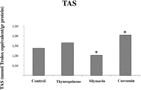
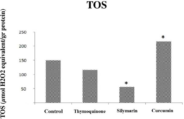
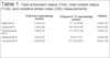
The effects of thymoquinone, silymarin, and curcumin on TAS, TOS, and OSI parameters are shown in Table 1. There was no difference in TAS values in the sham and control groups, but there was a significant difference in TOS and OSI values (P<0.05). The oxidant capacity was higher in the control group than in the sham group (Table 1). OSI values were found to be low in the thymoquinone group (P=0.009) (Figure 3), while there was no difference between the thymoquinone group and the control group in terms of TAS and TOS values (P>0.05) (P=0.175, P=0.347) (Figures 1 and 2). TAS, TOS, and OSI were found to be low in the silymarin-treated group when this group and the control group were compared (P=0.009) (Figures 1, 2, and 3). The levels of TAS and TOS were higher in the curcumin group than in the control group (P<0.05) (Figures 1 and 2) and OSI values were similar (Figure 3), unlike thymoquinone and silymarin groups. Regarding TAS values, there was no change in the thymoquinone group compared to the control group, whereas there was a 27% decrease in the silymarin group and a 48% increase in the curcumin group. Regarding TOS values, there was no change in the thymoquinone group compared to the control group, whereas there was a 63% decrease in the silymarin group and a 45% increase in the curcumin group. And regarding OSI values, there was a 33% decrease in the thymoquinone group, 49% decrease in the silymarin group, and no change in the curcumin group compared to the control group.


| TAS (Trolox equivalent/gr protein) |
TOS (μmol H 2O2 equivalent/gr protein) |
OSI (AU) |
|
|---|---|---|---|
| Sham (N=5) | 1.47±0.14 | 77.11±10.08 | 5.22±0.36 |
| Control (N=5) | 1.39±0.08 | 150.16±19.96# | 10.63±0.90# |
| Thymoquinone (N=5) | 1.66±0.16 | 117.12±11.09 | 7.09±0.25* |
| Silymarin (N=5) | 1.02±0.07** | 55.81±7.72** | 5.45±0.68** |
| Curcumin (N=5) | 2.05±0.09*** | 217.23±32.01*** | 10.62±1.46 |
DISCUSSION
One of the most important problems in the I-R mechanism is the total reactive oxygen products and the total antioxidant capacity against it[3-5]. There are many methods to measure oxidant and antioxidant capacity. Among these, TAS and TOS are easy, reliable, sensitive, and inexpensive methods that can be performed fully automatically for the measurement of total oxidant and antioxidant capacities[27,28]. Therefore, all three parameters showing oxidant and antioxidant capacities were quickly, easily, and reliably studied in our study. As a result of these evaluations, the oxidant capacity was found to be higher in the ischemia-treated control group compared to the sham group, which was to be expected. A significant difference was observed on the OSI value in the thymoquinone group compared to the control group (even though a decrease in TOS values was observed, no significant statistical difference was observed). This result indicates that thymoquinone activates antioxidant systems in I-R injury. This effect is similar to the presented in the study of Gökçe et al.[29], showing that thymoquinone reduces TOS and OSI values in I-R injury in rat testicular tissue. Thymoquinone probably performs its antioxidant activity by increasing antioxidant enzyme activities (SOD, CAT, GPx) and GSH levels[30]. Thymoquinone can also reduce the inflammatory activity that occurs with I-R. This demonstrates its efficacy by reducing inflammatory cytokines and reducing tumor necrosis factor-α[31]. This anti-inflammatory activity caused by thymoquinone causes a decrease in the amount of ROS formed.
Silymarin was found to be the herbal treatment that showed the best antioxidant activity in our study. Silymarin, which significantly reduced all the TAS, TOS, and OSI values, was thought to have better antioxidant properties than thymoquinone and curcumin. Silymarin has been used mainly in the treatment of liver and gastrointestinal diseases and is still used today against cirrhosis, chronic hepatitis, alcohol-related liver diseases, and various environmental toxic substances[24,32,33]. Two main mechanisms are proposed to explain the hepatoprotective property of silymarin. The first is based on its antioxidant effect due to its strong free radical scavenger, ROS, and lipid peroxidation reducing properties. The second is its anti-inflammatory and antiapoptotic mechanisms due to NF-κB modulation, NO modulation, and a decrease in COX-2 expression[20,32]. GSH levels have also been reported to increase similarly to thymoquinone[34]. Curcumin was found to reduce oxidative stress in rat ovary[35], prevent histopathological damage in mesenteric I-R injury and intestinal tissue, reduce lung damage, and reduce malondialdehyde, and other oxidative stress parameters[25,36]. It reduces endothelial dysfunction[37], myocardial I-R injury[38], and acts as a cardioprotective antioxidant[39,40]. Curcumin exhibits anti-inflammatory activity similarly to thymoquinone[22] and inhibits NF-κB metabolism similarly to silymarin[41]. Its efficacy in our study lagged behind the other two treatments even though curcumin was recognized as an effective antioxidant. We think that DMSO, which is used as a solvent in this effect of curcumin, plays a role as both prooxidant and antioxidant[42,43].
Distal organ damage was tried to be determined by gastrocnemius muscle histopathology. There were no histopathological changes in gastrocnemius muscle tissue for all groups in our study. We think that this is due to ischemia and reperfusion times and the insensitivity of gastrocnemius muscle tissue to I-R damage (muscle tissue is more resistant to I-R damage compared to other tissues). It was reported that cell death would not be seen by light microscopy until 10-12 hours after complete ischemia[44]. This supports our idea that the absence of histopathological change is due to low I-R times.
There are many studies related to herbal treatment methods and antioxidant studies when the literature is examined. However, the number of studies comparing these treatments is fairly limited. Therefore, our article will guide the extent to which the antioxidant activity of I-R after clamping of the abdominal aorta changes with thymoquinone, silymarin, and curcumin.
Limitations
This study has several limitations. The first relates to the administration of medications. Thymoquinone, silymarin, and curcumin were administered intraperitoneally. It is not foreseen how the effects of administering it in different ways (intravenous, oral) will be and how it will affect the effectiveness. The second is the effect of medication solvents in the study on oxidant and antioxidant processes. Third is that basic biochemical parameters and especially inflammatory parameters are not studied due to the anti-inflammatory efficacy of all three treatments. And the fourth and last limitation is that histopathological samples are not taken from different tissues, and changes in other tissues are not seen.
CONCLUSION
Thymoquinone and silymarin significantly reduce oxidative stress in the I-R injury model applied to the abdominal aorta. Silymarin’s antioxidant activity is much more effective compared to the other two agents. Curcumin’s antioxidant effect is much lower compared to the other two agents. Histopathological changes in peripheral end organ damage are thought to occur after longer I-R periods. Further studies are needed in the future to better understand the effects of thymoquinone, silymarin, and curcumin.
REFERENCES
1. De Grooth H., Rauen U. Ischemia-Reperfusion Injury: Processes in Pathogenetic Networks: A Review. Transplant Proc. 2007 Mar;39(2):481-4. doi: 10.1016/j.transproceed.2006.12.012.
2. Camara-Lemarroy C.R. Remote ischemic preconditioning as treatment for non-ischemic gastrointestinal disorders: Beyond ischemia-reperfusion injury. World J Gastroenterol. 2014 Apr 7;20(13):3572-81. doi: 10.3748/wjg.v20.i13.3572.
3. McMichael M., Moore R.M. Ischemia reperfusion injury pathophysiology, part I. Journal of Veterinary Emergency and Critical Care. 2004;14(4):231-241. 8.
4. Eltzschig H.K., Collard C.D. Vascular ischaemia and reperfusion injury. Br Med Bull. 2004 Oct 19;70:71-86. doi: 10.1093/bmb/ldh025. Print 2004. [MedLine]
5. Siemionow M., Arslan E. Ischemia/Reperfusion Injury: A Review In Relation To Free Tissue Transfers. Microsurgery. 2004;24(6):468-75. doi: 10.1002/micr.20060.
6. Abdollahi, M., Ranjbar, A., Shadnia, S., et al. Pesticides and oxidative stress: a review. Med Sci Monit. 2004 Jun;10(6):RA141-7.Epub 2004 Jun 1.
7. El-Gendy K.S., Aly N.M., Mahmoud F.H., et al. The role of vitamin C as antioxidant in protection of oxidative stress induced by imidacloprid. Food Chem Toxicol. 2010 Jan;48(1):215-21. doi: 10.1016/j.fct.2009.10.003. Epub 2009 Oct 13.
8. Sinha M., Manna P., Sil P.C.. Induction of necrosis in cadmium-induced hepatic oxidative stress and its prevention by the prophylactic properties of taurine. J Trace Elem Med Biol. 2009;23(4):300-13. doi: 10.1016/j.jtemb.2009.03.010. Epub 2009 May 6.
9. Dringen R. Metabolism and functions of glutathione in brain. Prog Neurobiol. 2000 Dec;62(6):649-71. doi: 10.1016/s0301-0082(99)00060-x.
10. Kulinsky V.I., Kolesnichenko L.S. The glutathione system. II. Other enzymes, thiol-disulfide metabolism, inflammation, and immunity, functions. Biomed Khim. Jul-Aug 2009;55(4):365-79.
11. Yener A.Ü., Çiçek M.C., Genç S.B., et al. Protective role of heparin in the injury of the liver and kidney on the experimental model of ischemia/reperfusion. J Cardiothorac Surg. 2014 Feb 17;9:35. doi: 10.1186/1749-8090-9-35.
12. Xiong J., Zhang M., Guo W., et al. Early malperfusion, ischemia reperfusion injury, and respiratory failure in acute complicated type B aortic dissection after thoracic endovascular repair. J Cardiothorac Surg. 2013 Jan 23;8:17. doi: 10.1186/1749-8090-8-17.
13. Pulathan Z., Altun G., Hemşinli D., et al. Role of Ethyl Pyruvate in Systemic Inflammatory Response and Lung Injury in an Experimental Model of Ruptured Abdominal Aortic Aneurysm. Biomed Res Int. 2014;2014:857109. doi: 10.1155/2014/857109. Epub 2014 Jan 19.
14. Jaeschke H., Woolbright B.L. Current strategies to minimize hepatic ischemia– reperfusion injury by targeting reactive oxygen species. Transplant Rev (Orlando). 2012 Apr;26(2):103-14. doi: 10.1016/j. trre.2011.10.006.
15. Hilal Alkis, Elif Demir, Mehmet Resit Taysi, et al. Effects of Nigella sativa oil and thymoquinone on radiation-induced oxidative stress in kidney tissue of rats. Biomed Pharmacother. 2021 Apr 5;139:111540. doi: 10.1016/j.biopha.2021.111540. Online ahead of print.
16. Bayrak O., Bavbek N., Karatas O.F., et al. Nigella sativa protects against ischaemia/reperfusion injury in rat kidneys. Nephrol Dial Transplant. 2008 Jul;23(7):2206-12. doi: 10.1093/ndt/gfm953. Epub 2008 Jan 22.
17. Ahmed M.A., Hassanein K.M.A. Cardio protective effects of Nigella sativa oil on lead induced cardio toxicity: Anti inflammatory and antioxidant mechanism. Journal of Physiology and Pathophysiology. 2013;4(5):72-80. doi: 10.5897/JPAP2013.0083
18. Khan A., Chen H., Tania M., et al. Anticancer Activities Of Nigella Sativa (Black Cumin). Afr J Tradit Complement Altern Med. 2011;8(5 Suppl):226-32. doi: 10.4314/ajtcam.v8i5S.10. Epub 2011 Jul 3.
19. Alessandro Federico, Marcello Dallio, Carmelina Loguercio. Silymarin/Silybin and Chronic Liver Disease: A Marriage of Many Years. Molecules. 2017 Jan 24;22(2):191. doi: 10.3390/ molecules22020191.
20. Shahbazi F., Khavidaki D.S., Khalili H., et al. Potential Renoprotective Effects of SilymarinAgainst Nephrotoxic Drugs: A Review of Literature. J Pharm Pharm Sci. 2012;15(1):112-23. doi: 10.18433/ j3f88s.
21. Raghavendhar R Kotha, Devanand L Luthria. Curcumin: Biological, Pharmaceutical, Nutraceutical, and Analytical Aspects. Molecules. 2019 Aug 13;24(16):2930. doi: 10.3390/molecules24162930.
22. Anthwal A., Thakur B.K., Rawat M.S.M., et al. Synthesis, characterization and in vitro anticancer activity of C-5 curcumin analogues with potential to inhibit TNF-alpha-induced NF-kappaB activation. BioMed Res. Int. 2014;2014:524161. doi: 10.1155/2014/524161. Epub 2014 Jul 24.
23. Mohamed A Kandeil, Safaa B Gomaa, Mohamed O Mahmoud. The effect of some natural antioxidants against cisplatin-induced neurotoxicity in rats: behavioral testing. Heliyon. 2020 Aug 26;6(8):e04708. doi: 10.1016/j.heliyon.2020.e04708. eCollection 2020 Aug.
24. Koçarslan A, Koçarslan S, Aydin MS, Gunay Ş, Karahan MA, Taşkın A, Üstunel M, Aksoy N. Intraperitoneal Administration of Silymarin Protects End Organs from Multivisceral Ischemia/ Reperfusion Injury in a Rat Model. Braz J Cardiovasc Surg. 2016 Nov-Dec;31(6):434-439. doi: 10.5935/1678-9741.20160072.
25. Mehmet Salih Aydin, Ahmet Caliskan, Aydemir Kocarslan, et al. Intraperitoneal curcumin decreased lung, renal and heart injury in abdominal aorta ischemia/reperfusion model in rat. Int J Surg. 2014;12(6):601-5. doi: 10.1016/j.ijsu.2014.04.013. Epub 2014 May 9
26. Elif Güneysu, Atacan Emre Koçman, Orhan Özatik, et al. The effects of iloprost and alprostadil on ischemia-reperfusion injury in preventing inflammation, tissue degeneration, and apoptosis in rat skeletal muscle. Turk J Med Sci. 2017 Jun 12;47(3):1028-1036. doi: 10.3906/sag-1604-59.
27. Erel O. A novel automated method to measure total antioxidant response against potent free radical reactions. Clin Biochem. 2004 Feb;37(2):112-9. doi: 10.1016/j.clinbiochem.2003.10.014.
28. O. Erel. A new automated colorimetric method for measuring total oxidant status. Clin Biochem. 2005 Dec;38(12):1103-11. doi: 10,1016/j.clinbiochem. 2005.08.008. Epub 2005 Oct 7.
29. Gökçe A., Oktar S., Koc A., et al. Protective effect of thymoquinone in experimental testicular torsion. Urol Int. 2010;85(4):461-5. doi: 10.1159/000318890. Epub 2010 Jul 13.
30. Tas U, Ayan M, Sogut E, et al. Protective effects of thymoquinone and melatonin on intestinal ischemia-reperfusion injury. Saudi J Gastroenterol. Sep-Oct 2015;21(5):284-9. doi: 10.4103/1319- 3767.166203.
31. Ali Parlar, Seyfullah Oktay Arslan. Thymoquinone reduces ischemia and re perfusion-induced intestinal injury in rats, through antioxidative and anti-inflammatory effects. Turk J Surg. 2020 Mar 18;36(1):96-104. doi: 10.5578/turkjsurg.4583. eCollection 2020 Mar.
32. Simin Dashti-Khavidaki, Foroud Shahbazi, Hossein Khalili, et al. Potential renoprotective effects of silymarin against nephrotoxic drugs: a review of literatüre. J Pharm Pharm Sci. 2012;15(1):112-23. doi: 10.18433/j3f88s.
34. Altaei T. Protective effect of silymarin during coronary artery bypass grafting surgery. Exp Clin Cardiol. 2012;17(1):34-38.
35. Sak M.E., Soydinc H.E., Sak S., et al. The protective effect of curcumin on ischemia-reperfusion injury in rat ovary. Int J Surg. 2013;11(9):967-70. doi: 10.1016/j.ijsu.2013.06.007. Epub 2013 Jun 21.
36. Onder A., Kapan M., Gümüş M, et al. The protective effects of curcumin on intestine and remote organs against mesenteric ischemia/reperfusion injury. Turk J Gastroenterol. 2012 Apr;23(2):141-7. doi: 10.4318/tjg.2012.0446.
37. Jeong G.S., Oh G.S., Pae H.-O., et al. Comparative effects of curcuminoids on endothelial heme oxygenase-1 expression: Ortho-methoxy groups are essential to enhance heme oxygenase activity and protection. Exp. Mol. Med. 2006 Aug 31;38(4):393-400. doi: 10.1038/emm.2006.46.
38. Cheol-Won Jeong, Kyung Yeon Yoo, Seong Heon Lee, et al. Curcumin protects against regional myocardial ischemia/ reperfusion injury through activation of RISK/GSK-3β and inhibition of p38 MAPK and JNK. J Cardiovasc Pharmacol Ther. 2012 Dec;17(4):387-94. doi: 10.1177/1074248412438102. Epub 2012 Mar 6.
39. Han J., Pan X.Y., Xu Y., Xiao Y., An Y., Tie L., et al. Curcumin induces autophagy to protect vascular endothelial cell survival from oxidative stress damage. Autophagy. 2012 May 1;8(5):812-25. doi: 10.4161/auto.19471. Epub 2012 May 1.
40. Yoona Kim, Peter Clifton. Curcumin, Cardiometabolic Health and Dementia. Int J Environ Res Public Health. 2018 Sep 24;15(10):2093. doi: 10.3390/ijerph15102093.
41. Fan Z., Jing H., Yao J., et al. The Protective Effects of Curcumin on Experimental Acute Liver Lesion Induced by Intestinal Ischemia-Reperfusion through Inhibiting the Pathway of NF-𝜅B in a rat Model. Oxid Med Cell Longev. 2014;2014:191624. doi: 10.1155/2014/191624. Epub 2014 Aug 0.
42. Min-Hee Kang, Joydeep Das, Sangiliyandi Gurunathan, et al. The cytotoxic effects of dimethyl sulfoxide in mouse preimplantation embryos: a mechanistic study. Theranostics. 2017 Oct 17;7(19):4735-4752. doi: 10.7150/thno.21662. eCollection 2017.
43. Carolina Sanmartín-Suárez, Ramón Soto-Otero, Inés Sánchez- Sellero, et al. Antioxidant properties of dimethyl sulfoxide and its viability as a solvent in the evaluation of neuroprotective antioxidants. J Pharmacol Toxicol Methods. Mar-Apr 2011;63(2):209-15. doi: 10.1016/j.vascn.2010.10.004. Epub 2010 Nov 6
44. Kumar V, Abbas AK, Fausto N, eds Robbins and Cotran Pathologic Basis of Disease, 7th Edition, Philadelphia: Elsevier Saunders, 2004.
33. Ting H., Deep G., Agarwal R. Molecular Mechanisms of Silibinin- Mediated Cancer Chemoprevention with Major Emphasis on Prostate Cancer. AAPS J. 2013 Jul;15(3):707-16. doi: 10.1208/ s12248-013-9486-2. Epub 2013 Apr 16.
Authors’Roles & Responsibilities
MY = Substantial contributions to the conception or design of the work; or the acquisition, analysis, or interpretation of data for the work; final approval of the version to be published
MG = Substantial contributions to the conception or design of the work; or the acquisition, analysis, or interpretation of data for the work; final approval of the version to be published
MSA = Substantial contributions to the conception or design of the work; or the acquisition, analysis, or interpretation of data for the work; final approval of the version to be published
NK = Substantial contributions to the conception or design of the work; or the acquisition, analysis, or interpretation of data for the work; final approval of the version to be published
ET = Substantial contributions to the conception or design of the work; or the acquisition, analysis, or interpretation of data for the work; final approval of the version to be published
Article receive on Monday, May 17, 2021
Article accepted on Wednesday, October 6, 2021
 All scientific articles published at rbccv.org.br are licensed under a Creative Commons license


All rights reserved 2017 / © 2024 Brazilian Society of Cardiovascular Surgery
DEVELOPMENT BY ![]()
 English PDF
 Print
Print
 Send this article by email
 How to cite this article
 Submit a comment
 Mendeley
 Pocket