![]()
![]()
Edward Araujo JúniorI; Luciane Alves da RochaII; Luciano Marcondes Machado NardozzaI
DOI: 10.5935/1678-9741.20140040
RESUMO
Doenças cardíacas congênitas são as malformações congênitas mais frequentes, entretanto, a detecção pré-natal ainda permanece baixa. A ecocardiografia bidimensional é o "padrão-ouro" para o rastreamento e diagnóstico das doenças cardíacas congênitas durante o pré-natal, entretanto, é operador dependente e realizada somente em gestantes de alto risco. Spatio-temporal image correlation é um software de ultrassonografia tridimensional que analisa o coração fetal e suas conexões vasculares nos modos multiplanar e superfície, contudo, também é operador dependente e consome muito tempo. Apresentamos um novo software: Sonocubic fine para o rastreamento das doenças cardíacas congênitas. Este software aplica a inteligência da tecnologia de navegação em volumes de spatio-temporal image correlation para automaticamente gerar nove planos ecocardiográficos padrões. Além disso, esta técnica tende a ser menos operador dependente e consumir menor tempo.
ABSTRACT
Congenital heart disease is the most common fetal congenital malformations; however, the prenatal rate detection still is low. The two-dimensional echocardiography is the "gold standard" exam to screening and diagnosis of congenital heart disease during the prenatal; however, this exam is operator-depending and it is realized only in high risk pregnancies. Spatio-temporal image correlation is a three-dimensional ultrasound software that analyses the fetal heart and your connections in the multiplanar and rendering modes; however, spatio-temporal image correlation too is operator-depending and time-consuming. We presenting a new three-dimensional software named Sonocubic fine to the screening of congenital heart disease. This software applies intelligent navigation technology to spatio-temporal image correlation volume datasets to automatically generate nine fetal echocardiography standard views. Thus, this new software tends to be less operator-depending and time-consuming.
AIUM: American Institute of Ultrasound in Medicine
CHD: Congenital heart disease
ISUOG: International Society of Ultrasound in Obstetrics & Gynecology
STIC: Spatio-temporal image correlation
INTRODUCTION
Congenital heart diseases (CHD) are the most common fetal congenital malformations, corresponding an incidence six times higher than chromosome anomalies and four times higher than neural tube defects[1]. The prevalence of CHD in newborns ranges from 0.6 to 5%[2]. The gold standard exam to the screening of CHD during the prenatal period is the two-dimensional echocardiography; however, despite the efforts realized in the last 20 years, the detection rate of CHD ranges from 31 to 96%[3].
Despite of two-dimensional echocardiography to be the gold standard exam to the screening of CHD during the prenatal period, this exam is operator dependent, is not available in all prenatal care centers and it is performed only in high risk pregnancies[4]. Due to this, some associations as International Society of Ultrasound in Obstetrics & Gynecology (ISUOG) and American Institute of Ultrasound in Medicine (AIUM) have proposed the screening of CHD in the second trimester scan including the outflow right and left ventricles tract to the four chamber view[5,6].
Spatio-temporal image correlation (STIC) is a software application that enables acquisition of fetal heart volume and vascular connection data. The images generated by this software can be evaluated both in multiplanar and rendering modes. They can also be evaluated both static and movement (4D) through a cineloop sequence that simulates an entire cardiac cycle[7-9]. Some studies have used the STIC in the screening of CHD; however, the results still are controversies[10-13]. In part, these controversy results are due of ability of examiners, being necessary specific training for long time to obtain the standard fetal heart views using STIC.
Recently, new software named Sonocubic fine (Medge Platforms Inc., New York, NY, USA) applies intelligent navigation technology to STIC volume datasets to automatically generate fetal echocardiography standard views. Thus, this new software tends to be less operator-depending and time-consuming.
The aim this study is to present the new software Sonocubic fine and its possible applications in screening of CHD.
METHODS
Technical description of the Sonocubic fine software: intelligent navigation; benefits of intelligent navigation; definition of Sonocubic fine; acquisition and storage of STIC volume datasets; anatomic box; intelligent alerts and virtual intelligent sonographer assistance (VIS-Assistance)
Intelligent navigation
Intelligent Navigation is referred to a completely unsupervised procedure able to find, extract and navigate target planes from a volume dataset using a predictive adapted algorithm. Any volume modality like ultrasound, computed tomographic, magnetic resonance imaging, could be used. The term "intelligent" describes the "operator independent" ability to solve a particular case by automated analysis of a number of variables leading to consequent adjustment of the predetermined imaging criteria.
Benefits of intelligent navigation
Once a correct volume is acquired (STIC), intelligent navigation has a role in obtaining the target diagnostic planes automatically, minimizing the intra-operator variability and the exam duration. The obtained planes are obtained following an expert criterion with a high rate of accuracy. The final images will be consistent in terms of orientation, sizes and positions simplifying the clinical evaluation of cases. Intelligent navigation identifies, isolates and independently process a sequence of selected particular morphology features of the anatomy in order to decide the more adequate plane to be extracted for that particular case.
As an example, the greater accuracy on the final image of a given cardiac volume is provided by the combination of anatomical probabilistic assumptions with corrective decision trees that increases the tolerance of the model to unprecedented range of fetal ages, position and anatomical variations. Intelligent navigation can label structures within the images for better interpretation.
Definition of Sonocubic fine
Sonocubic fine program has an intelligent navigation. The algorithm processes the volume data using some user marked reference points. These points, or landmarks, are marked on the original 2D sweep. The landmarks are specific anatomical spots sufficient for the algorithm to perform the volume navigation and deliver the diagnostic planes.
Acquisition and storage of STIC volume datasets
The volume to be available by Sonocubic fine should be acquired by STIC from Voluson 730 Expert or E8 (General Eletric, Medical System, Zipf, Austria) ultrasound machine using a convex volumetric transductor (RAB 4-8L). The best volume is obtained in four chamber view with the fetus spine in 6 o' clock position. The opening angle varies in according the gestational age, rather 20º to 25º in the second and 35º to 40º in the third trimesters. The acquisition time varied between 7.5 and 15 seconds, depending of fetus movements[14]. The volumes should be acquired only in gray scale. To storage of STIC volume datasets, we do not need to use the standardization proposed by Paladini[15] for fetuses with pelvic presentation, because the system automatically recognizes this situation and it requests to examiner to modify the fetus presentation.
Anatomic box
Before to active the anatomic box, the examiner should to click in the center of thoracic aorta in the four chamber view to observe the position of descendent aorta in the sagittal view. If the descendent aorta has linear direction, the STIC volume is appropriate to be analyzed by Sonocubic fine. Anatomic box is a technology allows the examiner to mark anatomical structures within the STIC volume (displayed as a cinellop - Video 1), which then allows geometrical modeling of the fetal heart and the automatic display of standard fetal echocardiography views. The seven landmarks structures to obtain the nine fetal echocardiography views are: 1) in the center of abdominal aorta in the upper abdomen; 2) center of thoracic aorta in the four chamber view; 3) heart crux in the four chamber view; 4) line crossing the middle of interventricular septum and reaching the internal wall of the right atrium in the four chamber view; 5) at level of pulmonary valve in three vessels and trachea view; 6) in the center of superior vena cava in the three vessels and trachea view; 7) in the transverse aortic arch in the three vessels and trachea view (Figure 1).

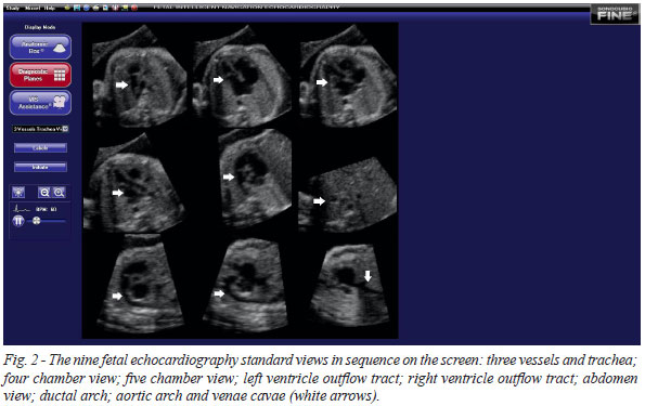
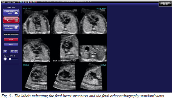
After the examiner to complete all landmarks, the program displays automatically the nine fetal echocardiography views: three vessels and trachea; four chamber view; five chamber view; left ventricle outflow tract; right ventricle outflow tract; abdomen view; ductal arch; aortic arch and venae cavae (Figure 2 and Video 2). The program permits too to add labels in each fetal echocardiography standard views, facilitating the correct understanding of fetal heart structure and the standard views (Figure 3). The examiner can make the adjustments of brightness, midtones and contrast; moreover, the fetal echocardiography views can be displayed in gray, sepia and ice colors.
In some cases, the program not shows the exact plane to examiner to mark the correct structures. In these cases, the examiner can make some adjustments using 2D plane position.
Intelligent Alerts
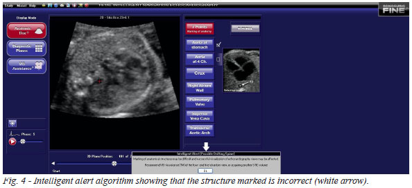
Intelligent alerts notify the examiner about the potential about potential issues with the STIC volume dataset (e.g. location of fetal spine in 8 o' clock) (Figure 4). Marking alerts are captions notifying the examiner that the fetal anatomical structures for making may in different locations that is expected.
Virtual Intelligent Sonographer Assistance
(VIS-Assistance)
VIS-Assistant is an operator-independent tool that allows sonographic navigation and exploration of surrounding structures in each of fetal echocardiography standard views (Video 3). This algorithm is the particular importance in suspected cases of CHD, because it permits to find the best plane in each fetal echocardiography views.
DISCUSSION
Limitations
Despite of potential benefits in the screening of CHD, some limitations are observed. The STIC volume should be scanned with the fetus spine between 7 and 8 o'clock position, because the Sonocubic fine software does not present the option of rotation of volume in the three orthogonal planes. This limitation can be time-consuming to scan the best volume in the four chamber view. The new software too does not have capacity to make measurements, which can be important in suspicion of some types of CHD. Other limitation of new software is not capacity to analyze STIC volumes with color Doppler; despite of the color Doppler is not part of guidelines of screening of CHD[5,6].
Advantages over STIC
Sonocubic fine is a software that display in the screen the nine fetal echocardiography standard views. Differently of STIC, which the standard planes are obtained by several reconstructions techniques[16,18], damaging the quality of the planes; Sonocubic fine provides direct views without artifacts transmission. Moreover, the Sonocubic fine is not operator-depending differently of STIC, not being necessary specific knowledge in fetal cardiology. Sonocubic fine is too less time-consuming, because it display automatically all fetal echocardiography standard views on the screen.
First experience with Sonocubic fine in the assessment of fetal heart
Recently, Yeo & Romero described the first experience of Sonocubic fine in the assessment of fetal heart. These authors tested the Sonocubic fine in 50 STIC volumes of normal cases and 4 STIC volumes of CHD (coarctation of aorta, tetralogy of Fallot, transposition of great vessels and pulmonary atresia with intact ventricular septum). They assessed the rate of visualization of fetal echocardiography views using diagnostic planes and/or VIS-Assistance. Sonocubic fine was able to generate nine fetal heart standard views using diagnostic planes in 78-100%, VIS-Assistance in 98-100% and a combination of diagnostic planes and/or VIS-Assistance in 98-100% of cases. In all cases of CHD, the new software demonstrated evidences of fetal heart abnormalities[19].
CONCLUSION
In summary, we presented a new software to the screening of CHD. Sonocubic fine permits the achievement of nine fetal echocardiography view using seven landmarks from STIC volume datasets. This software is less time and operator-depending than STIC; however, future studies comparing both techniques are necessary to prove the real applicability of Sonocubic fine in the screening of CHD.
Supplementary materials
Video 1. STIC volume dataset in cinellop sequence making an axial scan over the fetal chest and upper abdomen.
Video 2. The nine fetal echocardiography standard views on the screen in the cinellop sequence.
Video 3. Virtual Intelligent Sonographer Assistance (VIS-Assistance) navigation and exploration of surrounding structures in the ductal arch view.
REFERÊNCIAS
1. Carvalho JS, Mavrides E, Shinebourne EA, Campbell S, Thilaganathan B. Improving the effectiveness of routine prenatal screening for major congenital heart defects. Heart. 2002;88(4):387-91. [MedLine]
2. Grandjean H, Larroque D, Levi S. The performance of routine ultrasonographic screening of pregnancies in the Eurofetus Study. Am J Obstet Gynecol. 1999;181(2):446-54. [MedLine]
3. Stümpflen I, Stümpflen A, Wimmer M, Bernaschek G. Effect of detailed echocardiography as part of routine prenatal ultrasonographic screening on detection of congenital heart disease. Lancet. 1996;348(9031):854-7. [MedLine]
4. Allan L. Prenatal diagnosis of structural cardiac defects. Am J Med Genet C Semin Med Genet. 2007;145C(1):73-6. [MedLine]
5. International Society of Ultrasound in Obstetrics & Gynecology. Cardiac screening examination of the fetus: guidelines for performing the 'basic' and 'extended basic' cardiac scan. Ultrasound Obstet Gynecol. 2006;27(1):107-13. [MedLine]
6. Fetal Echocardiography Task Force; American Institute of Ultrasound in Medicine Clinical Standards Committee; American College of Obstetricians and Gynecologists; Society for Maternal-Fetal Medicine. AIUM practice guideline for the performance of fetal echocardiography. J Ultrasound Med. 2011;30(1):127-36. [MedLine]
7. Gonçalves LF, Lee W, Chaiworapongsa T, Espinoza J, Schoen ML, Falkensammer P, et al. Four-dimensional ultrasonography of the fetal heart with spatiotemporal image correlation. Am J Obstet Gynecol 2003;189(6):1792-802. [MedLine]
8. DeVore GR, Falkensammer P, Sklansky MS, Platt LD. Spatio-temporal image correlation (STIC): new technology for evaluation of the fetal heart. Ultrasound Obstet Gynecol. 2003;22(4):380-7. [MedLine]
9. Araujo Júnior E, Rolo LC, Simioni C, Nardozza LM, Rocha LA, Martins WP, et al. Comparison between multiplanar and rendering modes in the assessment of fetal atrioventricular valve areas by 3D/4D ultrasonography. Rev Bras Cir Cardiovasc. 2012;27(3):472-6. [MedLine] Visualizar artigo
10. Viñals F, Poblete P, Giuliano A. Spatio-temporal image correlation (STIC): a new tool for the prenatal screening of congenital heart defects. Ultrasound Obstet Gynecol. 2003;22(4):388-94. [MedLine]
11. Wanitpongpan P, Kanagawa T, Kinugasa Y, Kimura T. Spatio-temporal image correlation (STIC) used by general obstetricians is marginally clinically effective compared to 2D fetal echocardiography scanning by experts. Prenat Diagn. 2008;28(10):923-8. [MedLine]
12. Cohen L, Mangers K, Platt L, Julien S, Gotteiner N, Dungan J, et al. Quality of 2- and 3-dimensional fast acquisition fetal cardiac imaging at 18 to 22 weeks: ramifications for screening. J Ultrasound Med. 2009;28(5):595-601. [MedLine]
13. Araujo Júnior E, Rolo LC, Nardozza LM, Moron AF. Fetal cardiac evaluation by 3D/4D ultrasonography (STIC): what is its real applicability in the diagnosis of congenital heart disease? Rev Bras Cardiovasc. 2013;28(1):III-V. Visualizar artigo
14. Gonçalves LF, Lee W, Espinoza J, Romero R. Examination of the fetal heart by four-dimensional (4D) ultrasound with spatio-temporal image correlation (STIC). Ultrasound Obstet Gynecol. 2006;27(3):336-48. [MedLine]
15. Paladini D. Standardization of on-screen fetal heart orientation prior to storage of spatio-temporal image correlation (STIC) volume datasets. Ultrasound Obstet Gynecol. 2007;29(6):605-11. [MedLine]
16. DeVore GR, Polanco B, Sklansky MS, Platt LD. The 'spin' technique: a new method for examination of the fetal outflow tracts using three-dimensional ultrasound. Ultrasound Obstet Gynecol. 2004;24(1):72-82. [MedLine]
17. Espinoza J, Kusanovic JP, Gonçalves LF, Nien JK, Hassan S, Lee W, et al. A novel algorithm for comprehensive fetal echocardiography using 4-dimensional ultrasonography and tomographic imaging. J Ultrasound Med. 2006;25(8):947-56. [MedLine]
18. Yeo L, Romero R. Fetal Intelligent Navigation Echocardiography (FINE): a novel method for rapid, simple, and automatic examination of the fetal heart. Ultrasound Obstet Gynecol. 2013;42(3):268-84. [MedLine]
No financial support.
Authors' roles & responsibilities
EAJ: Proposal, preparation and article writing
LAR: Learning and collection of cases
LMMN: Critical review
Article receive on segunda-feira, 26 de agosto de 2013
 All scientific articles published at rbccv.org.br are licensed under a Creative Commons license


All rights reserved 2017 / © 2025 Brazilian Society of Cardiovascular Surgery
DEVELOPMENT BY ![]()
 Read in English
 English PDF
 Videos
 Print
Print
 Send this article by email
 How to cite this article
 Submit a comment
 Mendeley
 Pocket


