![]()
![]()
Fatih AksoyI; Serdar GulerI; Fatih KahramanI; Mevlüt Serdar KuyumcuI; Ali BagcıI; Hasan Aydın BasI; Dinçer UysalII; Ercan VarolI
DOI: 10.21470/1678-9741-2019-0062
ABSTRACT
Introduction: Metabolic syndrome (MetS) is defined as an association between diabetes, hypertension, obesity and dyslipidemia and an increased risk of cardiovascular disease. Mitral annular calcification (MAC) is associated with several cardiovascular disorders, including coronary artery disease, atrial fibrillation (AF), heart failure, ischemic stroke and increased mortality. The CHA2DS2-VASc score is used to estimate thromboembolic risk in AF. However, the association among MAC, MetS and thromboembolic risk is unknown and was evaluated in the current study.AF = Atrial fibrillation
BMP = Bone morphogenic protein
CI = Confidence interval
CVA = Cerebrovascular accident
HDL = High-density lipoprotein
IQR = Interquartile range
IVS = Interventricular septum
LA = Left atrial
LVEF = Left ventricular ejection fraction
LVEDD = Left ventricular end-diastolic diameter
LVESD = Left ventricular end-systolic diameter
LVPW = Left ventricular posterior wall
MAC = Mitral annular calcification
MetS = Metabolic syndrome
MESA = Multi-Ethnic Study of Atherosclerosis
OR = Odds ratio
OS = Oxidative stress
ROC = Receiver operating characteristics
TIA = Transient ischemic attack
INTRODUCTION
Mitral annular calcification (MAC) is a chronic, age-related, non-inflammatory degenerative process affecting the fibrous annulus of the mitral valve[1-3]. Recent studies demonstrated the strong association between the coexistence of MAC and cardiovascular risk factors[1,2]. In addition, studies have shown a relationship between MAC and carotid and peripheral artery disease[1,4]. MAC is accompanied by mitral valve disease and arrhythmias, including atrial fibrillation (AF) and conduction system disease and influenced the outcome of cardiac surgery[5,6]. Although it is most commonly asymptomatic and an incidental finding, its clinical relevance comes from the association of MAC with an increased rate of mortality and cardiovascular disease[7]. Additionally, a strong correlation was shown between MAC and risk of stroke[8]. Coexistence of MAC and stroke can be explained by cardiovascular risk factors, mitral valve disease and arrhythmias, including AF.
Metabolic syndrome (MetS) is a cluster of conditions including metabolic abnormalities, abdominal obesity, high blood pressure, hyperglycemia, low high-density lipoprotein (HDL) cholesterol levels, and high triglyceride levels. Additionally, patients with MetS have higher risk for heart disease, stroke and diabetes[9].
The CHA2DS2-VASc risk score is an inexpensive and easy scoring system that is calculated by assigning a score of 1 point for each of the following conditions: congestive heart failure (ejection fraction <40%), hypertension, age between 65 and 74 years, diabetes mellitus, vascular disease (myocardial infarction or peripheral arterial disease) and female gender; a score of 2 points for the following conditions: history of stroke or transient ischemic attack (TIA) and age >75 years. The CHA2DS2-VASc risk score is used to predict the risk of thromboembolism in patients with non-valvular AF[10].
In this study, we aimed to investigate the relationship between MAC, MetS and thromboembolic risk.
METHODS
Study Population
The 164 consecutive patients (78 females, 86 males, mean age 70.6±6.3 years) admitted to Suleyman Demirel University Hospital, Department of Cardiology outpatient clinic, and referred to our echocardiography laboratory due to suspicion of heart disease between June 2015 and May 2016 were enrolled in this prospective study. All patients underwent medical history, physical examination, anthropometric measurements, electrocardiogram, and echocardiographic evaluation. The study was approved by the institutional ethics committee and all patients gave their informed consent. Exclusion criteria were pericardial effusion, poor echocardiographic window, and history of chronic renal and liver disease, moderate to severe mitral and aortic regurgitation, moderate to severe mitral and aortic stenosis, malignancy, systemic or pulmonary embolism, chronic hematological diseases, acute or chronic inflammatory disease, autoimmune disease, hyperparathyroidism, hypercalcemia, hyperphosphatemia and presence of prosthetic valve. Patients were divided into two groups, according to the presence of MAC in echocardiographic evaluation.
Echocardiography
The M-mode, 2D and Doppler echocardiographic examinations were obtained by GE VingMed System FiVe (Norway) to assess left atrial (LA) diameter, interventricular septum (IVS) thickness, left ventricular posterior wall (LVPW) thickness, left ventricular-end diastolic diameter (LVEDD), left ventricular end-systolic diameter (LVESD), left ventricular ejection fraction (LVEF) and MAC. Left atrial and ventricular dimensions and LVEF were measured by M-mode echocardiography in the parasternal long axis view using the American Echocardiography Society M-mode technique[11]. MAC was defined as an intense echocardiographic-producing structure with highly reflective characteristics that was located at the junction of the atrioventricular groove and the posterior or anterior mitral leaflet on the parasternal long-axis, apical 4-chamber or 2-chamber, or parasternal short-axis view[12]. The presence of mitral and aortic insufficiency was evaluated by Doppler color flow mapping.
Metabolic Syndrome
At inclusion, MetS was defined according to the National Cholesterol Education Program Adult Treatment Panel III (NCEP ATP III) criteria as ≥3 of the following: (i) waist circumference ≥88 cm in women and ≥102 cm in men; (ii) elevated plasma triglycerides (≥1.7 mmol/L), or treatment for high triglycerides; (iii) low-plasma HDL-C (<1.3 mmol/L for women and <1.04 mmol/L for men); (iv) high fasting plasma glucose (≥5.6 mmol/L), or diabetes treatment; and (v) blood pressure ≥130/80 mmHg, or treated hypertension[13].
Statistical Analysis
SPSS version 16.0 software package was used for statistical analyses in this study. Categorical variables were expressed as frequency (%) and compared with the χ2 test. Kolmogorov-Smirnov test was used to test the distribution of numerical variables; those with normal distribution were expressed as mean ± standard deviation and were compared with Student’s t-test. Data without normal distribution were expressed as median [interquartile range (IQR) of 25%-75% percentiles] and were compared with the Mann-Whitney U test. In all statistical analyses, P-value <0.05 was considered statistically significant. The correlations between CHA2DS2-VASc risk score, presence of MetS, presence of MAC and other clinical, laboratory and echocardiographic parameters were performed with Pearson or Spearman correlation analysis when appropriate. Univariate analysis of binary logistic regression was carried out to identify which factors were associated with the presence of MAC. After including each of these potential confounding factors, backward conditional binary logistic regression analysis was performed to estimate the odds ratio (OR) and 95% confidence interval (95% CI) for presence of MAC. Receiver operating characteristics (ROC) curve analysis was used to analyze the prognostic value of CHA2DS2-VASc score for presence of MAC. C-Statistic (area under the curve) was presented as a unified estimate of sensitivity and specificity.
RESULTS
A total of 164 patients (mean age: 70.6 ± 6.3 years; range 52-85 years) were included in this study. In the echocardiographic evaluation, MAC was diagnosed in 94 patients. Demographic, clinical, laboratory and echocardiographic characteristics of the patients with and without MAC are listed in Table 1. Patients with MAC were generally older, more diabetic and more hypertensive when compared to patients without MAC (P<0.001, P<0.001 and P=0.001, respectively).
| MAC (-) (n=70) | MAC (+) (n=94) | P-value | |
|---|---|---|---|
| Age, years | 68.4±4.5 | 72.2±7.0 | <0.001 |
| Female gender, n (%) | 30 (42) | 45 (51) | 0.189 |
| Hypertension, n (%) | 29 (41) | 66 (70) | <0.001 |
| Diabetes mellitus, n (%) | 17 (24) | 47 (50) | 0.001 |
| Hyperlipidemia, n (%) | 33 (47) | 45 (47) | 0.526 |
| Smoking, n (%) | 19 (27) | 42 (44) | 0.016 |
| CAD, n (%) | 8 (11) | 23 (24.5) | 0.026 |
| CVA/TIA, n (%) | 1 (1.4) | 12 (12.8) | 0.006 |
| BMI (kg/m2) | 29.0±5.1 | 30.9±7.2 | 0.06 |
| Waist circumference (cm) | 97.4±12.4 | 101.0±11.0 | 0.05 |
| SBP (mmHg) | 118.0±17 | 121.0±17 | 0.182 |
| DBP (mmHg) | 75.0±8.1 | 77.0±10.3 | 0.09 |
| Heart rate (beat/min) | 70.1±12.4 | 71.7± 13.6 | 0.41 |
| Statin, n (%) | 16 (22) | 25 (26) | 0.359 |
| ACEi, n (%) | 13 (18) | 25 (26) | 0.154 |
| ARB, n (%) | 14 (20) | 24 (25) | 0.261 |
| OAD, n (%) | 7 (10) | 32 (34) | <0.001 |
| Insulin, n (%) | 4 (5.7) | 11 (11.7) | 0.149 |
| MetS, n (%) | 26 (37) | 60 (63) | 0.001 |
| CHA2DS2-VASc score | 2.1±1.3 | 3.5±1.3 | <0.001 |
| High risk group for CHA2DS2-VASc score | 42 (60) | 86 (91) | <0.001 |
| Platelet count (× 103/µL) | 248±81 | 242±61 | 0.58 |
| White blood cell count (× 103/µL) | 8.032±2.137 | 7.775±2.618 | 0.50 |
| HDL-cholesterol, mg/dL | 47.1±15.9 | 47.1±11.9 | 0.99 |
| LDL-cholesterol, mg/dL | 108.4±38.8 | 113.6±38.3 | 0.40 |
| Triglycerides, mg/dL | 154.1±119.22 | 147.9±75.3 | 0.70 |
| Glucose, mg/dL | 122.6±58.6 | 123.9±57.6 | 0.88 |
| Creatinine, mg/dL) | 0.9 ±0.1 | 0.9 ± 0.1 | 0.62 |
| Total cholesterol, mg/dL | 184.7±57.5 | 190±47.4 | 0.47 |
| LVEF, % | 60 ±2.2 | 58.7±4.1 | 0.02 |
| Aorta (mm) | 24.9 ±2.3 | 25.9±2.3 | 0.005 |
| LA (mm) | 34.7±4.8 | 37.4±4.3 | <0.001 |
| IVS (mm) | 10.5±1.3 | 11.2±1.4 | 0.001 |
| LVPW (mm) | 9.6±0.8 | 10±0.8 | 0.012 |
| LVESD (mm) | 27±2.3 | 28±3.3 | <0.001 |
| LVEDD (mm) | 44.6±2.5 | 46.2±3.2 | 0.001 |
ACEi=angiotensin converting enzyme inhibitor; ARB=angiotensin receptor blocker; BMI=body mass index; CAD=coronary artery disease; DBP=diastolic blood pressure; HDL=High density lipoprotein; IVS=interventricular septum; LA=left atrium; LDL=low density lipoprotein; LVEDD=left ventricular end diastolic diameter; LVEF=left ventricular ejection fraction; LVESD=left ventricular end systolic diameter; LVPW=left ventricular posterior wall; MAC=mitral annular calcification; MetS=metabolic syndrome; OAD=oral antidiabetics; SBP=systolic blood pressure; TIA=transient ischemic attack

Smoking, coronary artery disease (CAD) and stroke/TIA rates were higher in patients with MAC than patients without MAC (P=0.016, P=0.026 and P=0.006, respectively). Hyperlipidemia and gender were similar between patients with and without MAC (for both parameters, P>0.05). MetS rates were higher in patients with MAC (P=0.001). Patients with MAC had higher CHA2DS2-VASc scores than patients without MAC (P<0.001). When, according to CHA2DS2-VASc score, patients were divided into two groups, with 0 and 1 scores considered as low risk, and ≥2 scores considered as high risk, patients with MAC had a higher rate of high risk category than patients without MAC (P<0.001). There was no statistically significant difference between two groups for cholesterol and glucose levels (for both parameters, P>0.05). LA diameter was significantly higher in patients with MAC than without MAC (37.4±4.3 vs. 34.7±4.8 mm, respectively; P<0.001). IVS thickness was significantly higher in patients with MAC than without MAC (11.2±1.4 vs. 10.5±1.3 mm, respectively; P=0.001). LVEDD was significantly higher in patients with MAC than without MAC (46.2±3.2 vs. 44.6±2 mm, respectively; P=0.001). LVESD was significantly higher in patients with MAC than without MAC (28±3.3 vs. 27±2.3 mm, respectively; P<0.001).
Binary Logistic Regression for MAC
Correlation analysis indicated that MAC was positively correlated with CHA2DS2-VASc score (P<0.001, r=0.49), AF (P=0.008, r=0.208), LA diameter (P=0.001, r=0.267), age (P<0.001, r=0.347), history of hypertension (P<0.001, r=0.288), history of diabetes mellitus (P=0.001, r=0.261), presence of MetS (P=0.001, r=0.264), history of smoking (P=0.021, r=0.179), history of cerebrovascular accident/transient ischemic attack (CVA/TIA) (P=0.008, r=0.208), presence of AF (P=0.008, r=0.208), and negatively correlated with LVEF (P=0.021, r= -0.18) (Table 2).
| CHA2DS2-VASc score | Age | Hypertension | Diabetes mellitus | LA diameter |
LVEF | MetS | Smoking | CVA/TIA | AF | ||
|---|---|---|---|---|---|---|---|---|---|---|---|
| MAC | r | 0.490 | 0.347 | 0.288 | 0.261 | 0.267 | -0.180 | 0.264 | 0.179 | 0.208 | 0.208 |
| P | <0.001 | <0.001 | <0.001 | 0.001 | 0.001 | 0.021 | 0.001 | 0.021 | 0.008 | 0.008 |

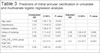
Univariate analysis showed that high CHA2DS2-VASc score and group, presence of MetS, advanced age, history of hypertension, history of diabetes mellitus and history of smoking were significantly associated with a higher risk of MAC (Table 3). A multivariate binary logistic regression analysis was carried out including all the characteristics associated with MAC in the univariate analysis. This analysis showed that high risk according to the CHA2DS2-VASc score (OR: 5.0; 95% CI: 2.0-12.5, P<0.001), presence of MetS (OR: 1.002; 95% CI: 1.00-1.003, P=0.014) and history of smoking (OR: 2.09; 95% CI: 1.00-4.35, P=0.049) remained as independent factors for MAC. ROC curve analysis showed that CHA2DS2-VASc score (C-statistic: 0.78; 95% CI: 0.706-0.855, P<0.001) were significant predictors of MAC (Figure 1). We calculated that a cut-off point of 2.5 for CHA2DS2-VASc scores could estimate the presence of MAC with a sensitivity of 81% and 70%. When univariate analysis was carried out for CVA/TIA, presence of MAC increased 10-fold the CVA/TIA risk (OR: 10, 09; 95% CI: 1.28-79.62, P=0.028).
| Unadjusted OR | CI 95% | P-value | Adjusted OR | CI 95% | P-value | |
|---|---|---|---|---|---|---|
| Age, years | 1.106 | 1.04-1.16 | <0.001 | |||
| Hypertension | 3.33 | 1.74-6.37 | <0.001 | |||
| Diabetes mellitus | 3.11 | 1.58-6.15 | 0.001 | |||
| History of smoking | 2.16 | 1.11-4.21 | 0.023 | 2.09 | 1.00-4.35 | 0.049 |
| MetS | 2.98 | 1.57-5.6 | 0.001 | 2.44 | 1.20-4.96 | 0.014 |
| CHA2DS2-VASc group (high risk) | 7.1 | 3.0-17.0 | <0.001 | 5.0 | 2.0-12.5 | <0.001 |
| CHA2DS2-VASc score | 2.26 | 1.68-3.0 | <0.001 |


DISCUSSION
The current study showed that higher CHA2DS2-VASc scores and presence of MetS were independently associated with the development of MAC; consequently, individuals at high risk according to CHA2DS2-VASc scores and presence of MetS should receive more attention to reduce unfavorable cardiovascular risk factors and the development of future cardiovascular events. The other finding of the study determined that the presence of MAC increased the risk of CVA/TIA and AF.
CHA2DS2-VASc score was associated with in-hospital and long-term adverse clinical outcomes, including mortality, in patients with stable CAD and acute coronary syndrome [14,15]. To the best of our knowledge, there is no study investigating the relationship between CHA2DS2-VASc score, MetS and MAC, which has similar risk factors for atherosclerotic heart disease.
Since MAC is often associate with coronary risk factors, such as hypertension, diabetes and obesity[2], it is possible that convergence of multiple risk factors could potentiate cardiovascular risk. This is exemplified by MetS. MAC is largely thought to be a manifestation of atherosclerotic coronary heart disease. This is mainly because the risk factors for MAC have been found to be similar to that of coronary artery disease, sharing a common pathogenesis for atherosclerosis.
MAC is associated with several cardiovascular disorders, including CAD, carotid and aortic atherosclerosis, heart failure, and stroke[2,4,8]. Previous studies have determined that age, hypertension, diabetes mellitus, and obesity, which are risk factors for atherosclerotic heart disease, are also risk factors for MAC[2,16]. In our study, we have found higher rates of diabetes mellitus and hypertension, which are a component of MetS and CHA2DS2-VASc score, in patients with MAC than in controls, and our results are consistent with previous studies of this aspect[2,16]. Bone morphogenic protein (BMP) 2, which is the key of osteogenic regulatory factor, is upregulated by hyperglycemia in vitro[17,18]. BMP2 showed to play a role in vascular calcification[17] and has been detected in areas of valvular calcification[18]. Another theory that explained increased calcium in mitral annulus is the pro-oxidative status in diabetes mellitus. Oxidative stress (OS) is caused by increased production of reactive oxygen species. Increased oxidative products lead to cell death or to acceleration in ageing and age-related disease[19]. Besides, OS not only plays a role in the pathogenesis of MAC and may explain increased mortality and morbidity in patients with MAC. OS is also known to regulate different stages of thrombotic processes, including platelet activation. OS may trigger platelet hyperactivity, reducing nitric oxide bioavailability[20]. Additionally, OS have a role in the metabolic syndrome process[21]. Thus, increasing OS may play a role in increased mortality and morbidity in patients with MAC.
Studies have demonstrated that MetS was associated with increased rates of aortic stenosis progression and bioprosthetic valve deterioration[22,23]. In Multi-Ethnic Study of Atherosclerosis (MESA) cohort, Katz et al.[24]showed that MetS was associated with new and older aortic valve. In our study, we demonstrated that patients with MetS had higher MAC rates than patients without MetS. In the MESA[5], investigators examined the association between MAC and AF in a racially and ethnically diverse population. They found that MAC was associated with an increased risk of AF (HR=1.9, 95% CI=1.5, 2.5). Hypertension and diabetes mellitus were more seen in patients with MAC than without MAC. Additionally, patients with MAC had left ventricular hypertrophy and left atrial enlargement more explicit than patients without MAC. Similarly, in the present study, we found that hypertension and diabetes mellitus were more observed in patients with MAC than without MAC. In addition, we have also found higher left atrial diameter, lower ejection fraction and higher incidence of left ventricular hypertrophy in patients with MAC than in controls. These findings may explain the association of MAC with adverse cardiovascular events, including stroke and increased mortality. Our other finding that AF and CVA/TIA were more seen in patients with MAC than without MAC, was consistent with left atrial dilatation and left ventricular hypertrophy. Our results are consistent with previous studies also in this respect[3,5].
The global prevalence of MetS has increased due to the increased rates of obesity and sedentary lifestyle and has been associated with a higher risk of major adverse cardiac events in the general population[25]. Individuals with MetS should receive more attention to reduce unfavorable cardiovascular risk factors and the development of future cardiovascular events. Additionally, lifestyle changes and cardiovascular risk modification may reduce cardiac structural changes, such as left atrial dilatation, left ventricular hypertrophy and MAC formation.
CONCLUSION
In conclusion, we showed that CHA2DS2-VASc score and presence of MetS rates were significantly higher in patients with MAC compared with controls. MAC was correlated with CHA2DS2-VASc score, presence of MetS, AF and left atrial diameter and negatively correlated with LVEF. The CHA2DS2-VASc score and presence of MetS were independently associated with presence of MAC. Presence of MetS and high CHA2DS2-VASc scores may indicate that patients with MAC have a higher risk of systemic thromboembolism.
REFERENCES
1. Adler Y, Fink N, Spector D, Wiser I, Sagie A. Mitral annuluscalcification-a window to diffuse atherosclerosis of the vascular system.Atherosclerosis. 2001;155(1):1-8.doi:10.1016/s0021-9150(00)00737-1. [MedLine]
2. Kanjanauthai S, Nasir K, Katz R, Rivera JJ, Takasu J, Blumenthal RS,et al. Relationships of mitral annular calcification to cardiovascular riskfactors: the multi-ethnic study of atherosclerosis (MESA). Atherosclerosis.2010;213(2):558-62. doi:10.1016/j.atherosclerosis.2010.08.072.
3. Ariyarajah V, Apiyasawat S, Barac I, Spodick DH. Is the presence ofmitral annular calcification associated with poor left atrial function?Echocardiography. 2009;26(8):877-84.doi:10.1111/j.1540-8175.2009.00900.x
4. Barasch E, Gottdiener JS, Larsen EKM, Chaves PH, Newman AB, ManolioTA. Clinical significance of calcification of the fibrous skeleton of the heartand aortosclerosis in community dwelling elderly. The cardiovascular healthstudy (CHS). Am Heart J. 2006;151(1):39-47.doi:10.1016/j.ahj.2005.03.052. [MedLine]
5. O’Neal WT, Efird JT, Nazarian S, Alonso A, Heckbert SR, Soliman EZ.Mitral annular calcification and incident atrial fibrillation in themulti-ethnic study of atherosclerosis. Europace. 2015;17(3):358-63.doi:10.1093/europace/euu265.
6. Okada Y. Surgical management of mitral annular calcification. GenThorac Cardiovasc Surg. 2013;61(11):619-25.doi:10.1007/s11748-013-0207-7.
7. Fox CS, Vasan RS, Parise H, Levy D, O'Donnell CJ, D'Agostino RB, etal. Mitral annular calcification predicts cardiovascular morbidity andmortality: the Framingham heart study. Circulation. 2003;107(11):1492-6.doi:10.1161/01.cir.0000058168.26163.bc.
8. De Marco M, Gerdts E, Casalnuovo G, Migliore T, Wachtell K, Boman K,et al. Mitral annular calcification and incident ischemic stroke in treatedhypertensive patients: the LIFE study. Am J Hypertens. 2013;26(4):567-73.doi:10.1093/ajh/hps082.
9. Mottillo S, Filion KB, Genest J, Joseph L, Pilote L, Poirier P, etal. The metabolic syndrome and cardiovascular risk a systematic review andmeta-analysis. J Am Coll Cardiol. 2010;56(14):1113-32.doi:10.1016/j.jacc.2010.05.034.
10. Kirchhof P, Benussi S, Kotecha D, Ahlsson A, Atar D, Casadei B, etal. 2016 ESC Guidelines for the management of atrial fibrillation developed incollaboration with EACTS. Eur Heart J. 2016;37(38):2893-962.doi:10.1093/eurheartj/ehw210. [MedLine]
11. Sahn DJ, DeMaria A, Kisslo J, Weyman A. Recommendations regardingquantitation in M-mode echocardiography: results of a survey ofechocardiographic measurements. Circulation. 1978;58(6):1072-83.doi:10.1161/01.cir.58.6.1072.
12. Kohsaka S, Jin Z, Rundek T, Boden-Albala B, Homma S, Sacco RL, etal. Impact of mitral annular calcification on cardiovascular events in amultiethnic community: the Northern Manhattan study. JACC Cardiovasc Imaging.2008;1(5):617-23. doi:10.1016/j.jcmg.2008.07.006.
13. Grundy SM, Cleeman JI, Daniels SR, Donato KA, Eckel RH, Franklin BA,et al. Diagnosis and management of the metabolic syndrome: an American heartassociation/national heart, lung, and blood institute scientific statement.Circulation. 2005;112(17):2735-52. Erratum in: Circulation. 2005;112(17):e297-8.doi:10.1161/CIRCULATIONAHA.105.169404.
14. Tasolar H, Çetin M, Balli M, Bayramoglu A, Otlu YÖ, Türkmen S, etal. CHA2DS2-VASc-HS score in non-ST elevation acute coronary syndrome patients:assessment of coronary artery disease severity and complexity and comparison toother scoring systems in the prediction of in-hospital major adversecardiovascular events. Anatol J Cardiol. 2016;16(10):742-8.doi:10.14744/AnatolJCardiol.2015.6593. [MedLine]
15. Capodanno D, Rossini R, Musumeci G, Lettieri C, Senni M, ValsecchiO, et al. Predictive accuracy of CHA2DS2-VASc and HAS-BLED scores in patientswithout atrial fibrillation undergoing percutaneous coronary intervention anddischarged on dual antiplatelet therapy. Int J Cardiol. 2015;199:319-25.doi:10.1016/j.ijcard.2015.07.064.
16. Boon A, Cheriex E, Lodder J, Kessels F. Cardiac valve calcification:characteristics of patients with calcification of the mitral annulus or aorticvalve. Heart. 1997;78(5):472-4. doi:10.1136/hrt.78.5.472.
17. Boström K, Watson K, Horn S, Wortham C, Herman I, Demer L. Bonemorphogenetic protein expression in human atherosclerotic lesions. J ClinInvest. 1993;91(4):1800-9. doi:10.1172/JCI116391.
18. Mohler ER 3rd, Gannon F, Reynolds C, Zimmerman R, Keane MG, KaplanFS. Bone formation and inflammation in cardiac valves. Circulation.2001;103(11):1522-8. doi:10.1161/01.cir.103.11.1522.
19. Kangralkar V, Patil SD, Bandivadekar R. Oxidative stress anddiabetes: a review. Int J Pharm Appl. 2010;1(1):38-45.
20. Fuentes E, Palomo I. Role of oxidative stress on platelethyperreactivity during aging. Life Sci. 2016;148:17-23.doi:10.1016/j.lfs.2016.02.026. [MedLine]
21. Francisqueti FV, Chiaverini LCT, Santos KCd, Minatel IO, Ronchi CB,Ferron AJT, et al. The role of oxidative stress on the pathophysiology ofmetabolic syndrome. Rev Assoc Med Bras. 2017;63(1):85-91.doi:10.1590/1806-9282.63.01.85.
22. Briand M, Lemieux I, Dumesnil JG, Mathieu P, Cartier A, Després JP,et al. Metabolic syndrome negatively influences disease progression andprognosis in aortic stenosis. J Am Coll Cardiol. 2006;47(11):2229-36.doi:10.1016/j.jacc.2005.12.073.
23. Briand M, Pibarot P, Després JP, Voisine P, Dumesnil JG, Dagenais F,et al. Metabolic syndrome is associated with faster degeneration ofbioprosthetic valves. Circulation. 2006; 114(1 Suppl):I512-7.doi:10.1161/CIRCULATIONAHA.105.000422.
24. Katz R, Budoff MJ, Takasu J, Shavelle DM, Bertoni A, Blumenthal RS,et al. Relationship of metabolic syndrome with incident aortic valve calcium andaortic valve calcium progression: the multi-ethnic study of atherosclerosis(MESA). Diabetes. 2009; 58(4):813-9. Erratum in: Diabetes. 2009;58(8):1937.doi:10.2337/db08-1515.
25. Grundy SM. Metabolic syndrome update. Trends Cardiovasc Med.2016;26(4):364-73. doi:10.1016/j.tcm.2015.10.004.
No financial support.
No conflict of interest.
Authors’ roles & responsibilities
FA Substantial contributions to the conception or design of the work; or the acquisition, analysis, or interpretation of data for the work; drafting the work or revising it critically for important intellectual content; final approval of the version to be published
SG Substantial contributions to the conception or design of the work; or the acquisition, analysis, or interpretation of data for the work; final approval of the version to be published
FK Substantial contributions to the conception or design of the work; or the acquisition, analysis, or interpretation of data for the work; drafting the work or revising it critically for important intellectual content; final approval of the version to be published
MSK Agreement to be accountable for all aspects of the work in ensuring that questions related to the accuracy or integrity of any part of the work are appropriately investigated and resolved
AB Substantial contributions to the conception or design of the work; or the acquisition, analysis, or interpretation of data for the work; drafting the work or revising it critically for important intellectual content; final approval of the version to be published
HAB Agreement to be accountable for all aspects of the work in ensuring that questions related to the accuracy or integrity of any part of the work are appropriately investigated and resolved
DU Drafting the work or revising it critically for important intellectual content
EV Substantial contributions to the conception or design of the work; or the acquisition, analysis, or interpretation of data for the work; drafting the work or revising it critically for important intellectual content; final approval of the version to be published
Article receive on Wednesday, February 13, 2019
Article accepted on Friday, March 1, 2019
 All scientific articles published at rbccv.org.br are licensed under a Creative Commons license


All rights reserved 2017 / © 2024 Brazilian Society of Cardiovascular Surgery
DEVELOPMENT BY ![]()
 English PDF
 Print
Print
 Send this article by email
 How to cite this article
 Submit a comment
 Mendeley
 Pocket