Surgical treatment of aortic dissection is a challenge for the cardiac surgeon, especially in patients undergoing cardiac operations. Our objective in this case report is to demonstrate how we treat the chronic type A aortic dissection in patients revascularized using percutaneous arterial and venous cannulae.
O tratamento operatório da dissecção aórtica é um desafio para o cirurgião cardíaco, sobretudo nos pacientes submetidos a operação cardíaca prévia. Nosso objetivo neste relato de caso é demonstrar como tratamos a dissecção aórtica crônica tipo A em paciente revascularizado utilizando cânulas arterial e venosa percutâneas.
INTRODUCTION
Hypertrophic cardiomyopathy (HCM) is an autosomal dominant genetic trait and is characterized by left ventricular hypertrophy, especially of the interventricular septum in the absence of other conditions that justify this anatomic change [1,2].
The sudden cardiac death (SCD), with an annual incidence of 1%, is the most serious complication of this disease [2,3]. The risk of fatal arrhythmias increases, however, when: the thickness of the ventricular septum is greater than 30 mm; ventricular tachycardia is detected, the patient reports syncope or when there are sudden deaths in young relatives. The prevention of SCD with the use of automatic cardioverter defibrillator (ICD) has been recommended in high risk patients [1-3].
There are few reports of ICD implantation in pregnant women and there is no standard procedure for this condition [4]. The application of shock therapy in pregnant women with IDC, reported in two studies, had no effect on fetal development [4,5].
The aim of this report is to describe the case of two patients with HCM who underwent ICD implantation for prevention of SCD in pregnancy intercourse.
CASE REPORTS
Case 1
JMG, 24 years old, at 29 weeks' gestation, presented syncope since nine months without prodrome or associated factors. A transthoracic echocardiogram reveale HCM with ventricular septum of 29 mm and 24-hour ECG detected non-sustained ventricular tachycardia (NSVT). The patient reported she had a brother with ICD due to recovered cardiorespiratory arrest and HCM.
Case 2
TIGC, 17 years old, at 26 weeks' gestation, she had repeated episodes of syncope without prodrome or triggering factors, associated with progressive dyspnea. Transthoracic echocardiography diagnosed HCM with interventricular septum with 30 mm thick. She complained of sudden death without etiologic diagnosis in young brother and mother recently diagnosed with HCM.
ICD implantation technique
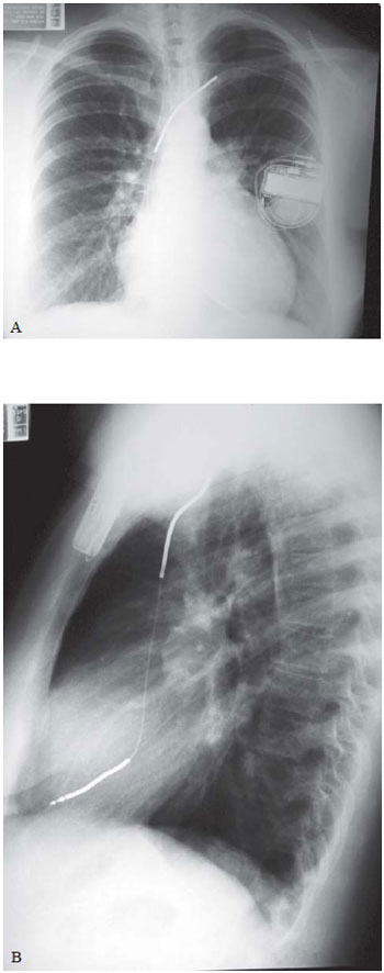
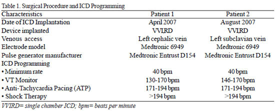
The implant procedure was performed in both cases, under intravenous sedation and local anesthesia. The abdomen of the patients was protected by blanket of lead. The transvenous lead was implanted with the aid of fluoroscopy, in the apical septum of the right ventricle and the pulse generator, housed in the left infraclavicular subcutaneous position (Figure 1). Defibrillation test was not performed.

Fig. 1 - A: Chest radiograph in the anteroposterior position (A) and lateral (B), showing the position of the electrode leads of the ICD implanted through the left cephalic vein. B: Chest radiograph in the anteroposterior position (A) and lateral (B), showing the position of the electrode leads of the ICD implanted through left cephalic vein

Both pregnancies were no abnormalities. During delivery, the heart rate was monitored and shock therapies were turned off, turning the previous program of ICD after the procedure.
At the end of puerperium, patients were tested with defibrillation. Under intravenous sedation, ventricular fibrillation was induced, and in both cases, the 20J automatic shock was effective to defibrillate the heart.
During follow-up of 2.7 ± 0.2 years, there were no reports of suggestive episodes of low cerebral blood flow or heart failure. The counters of diagnoses of the ICD did not record appropriate therapy for tachycardia or ventricular fibrillation. One patient presented, however, inappropriate shock therapies for murmur detection and required replacement of the lead, which showed increased pacing impedance. There were no deficiencies or growth disorders in children of these pregnancies.
DISCUSSION
Despite its low prevalence, HCM is an important cause of SCD in young individuals [1-6]. The main risk factors for fatal arrhythmias in these patients are a history of sudden death in young relatives, the presence of syncope of unknown origin, hypotension during exercise, ventricular septal thickness of e" 30 mm and episodes of NSVT on Holter of 24 hours [1-3,6].
In this report, the diagnosis of HCM was performed during pregnancy. The identification of multiple risk factors for MSC justified the indication of using ICD during pregnancy.
In addition to the usual anesthetic care for pregnant women, two points deserve special attention: the risk of malformations by use of fluoroscopy used to guide the placement of electrode leads and the lack of knowledge of the consequences of induction of ventricular fibrillation and shock application, that are necessary to test the integrity and efficiency of the system deployed.
Among the alternatives described to prevent fetal exposure to radiation, it has been proposed the implant guided by echocardiography [7] or by electroanatomic mapping [8]. In this report, given the low risk of fetal damage from radiation in the third trimester of pregnancy, we chose to fluoroscopy, associated with the use of lead blanket on the abdomen of the pregnant women, as additional protection.
There is no evidence in the literature that the induction of ventricular fibrillation and the shocks applied to the defibrillator test during implantation cause fetal abnormalities.
The automatic application of shock in pregnant women already bearer of ICD did not cause maternal or fetal effects [4,5]. Moreover, the real need for intraoperative defibrillation test is object of controversy. Clinical studies show that in only 4% of patients shocks are ineffective in reversing ventricular fibrillation induced during ICD implantation [9,10]. These studies also showed that ineffective shocks are infrequent in patients with preserved ventricular contractility [9,10]. In the cases reported herein, we opted for no defibrillation testing.
The low risk of malformations by radiation after the first trimester of pregnancy, which allowed the use of fluoroscopy, and good ventricular function of patients which avoided the test of defibrillation, became the ICD implantation in these patients a routine procedure, despite the current pregnancy.
1. Still RJ, Hilgenberg AD, Akins CW, Daggett WM, Buckley MJ. Intraoperative aortic dissection. Ann Thorac Surg. 1992;53(3):374-9.
2. Chavanon O, Carrier M, Cartier R, Hébert Y, Pellerin M, Pagé P, et al. Increased incidence of acute ascending aortic dissection with off-pump aortocoronary bypass surgery? Ann Thorac Surg. 2001;71(1):117-21. [MedLine]
3. Bopp P, Perrenoud JJ, Périat M. Dissection of ascending aorta. Rare complication of aortocoronary venous bypass surgery. Br Heart J. 1981:46(5):571-3. [MedLine]
4. Nicholson WJ, Crawley IS, Logue RB, Dorney ER, Cobbs BW, Hatcher CR Jr. Aortic root dissection complicating coronary bypass surgery. Am J Cardiol. 1978;41(1):103-7. [MedLine]
5. Hagl C, Ergin MA, Galla JD, Spielvogel D, Lansman S, Squitieri RP, et al. Delayed chronic type A dissection following CABG: implications for evolving techniques of revascularization. J Card Surg. 2000;15(5):362-7. [MedLine]


 All scientific articles published at rbccv.org.br are licensed under a Creative Commons license