ABSTRACT
Objective: The aim of this study was to assess clinical complications of limbs undergone harvesting of the great saphenous vein for venous coronary artery bypass graft surgery using bridge technique. Methods: Fourty-four patients who had undergone CABG using the great saphenous vein harvested by the bridge technique over more than 3 months ago were randomly selected. The exclusion criteria were the harvesting of both saphenous veins, prior saphenectomy of the contralateral limb, edema caused by a systemic etiology, such as heart, renal, thyroid or hepatic diseases and venous insufficiency of the lower limbs as characterized by swollen varicose veins both with and without trophic changes. The age, gender, diabetes, time of surgery and occurrence of complications, such as edema, paresthesia, infection, lymphorrhea, erysipelas and deep venous thrombosis, were assessed. The assessment was clinic and diagnosis of the diabetes was performed by the preoperative exams. The chi-square, Fisher and Student's t tests were used for statistical analysis with an alpha error of 5%. Results: The time between surgery and assessment ranged between 3 and 187 months with a mean of 47.3±42.5 months. Infections of the saphenous harvest site were detected in 25% of the cases, edema in 52.3%, paresthesia in 29.5%, erysipelas in 9.1%, lymphorrhea in 4.5% and deep venous thrombosis in 2.3%. There was no association between diabetes and complications. Conclusion: The saphenous vein harvesting using bridge technique for coronary artery bypass grafting does not eliminate clinical complications, such as paresthesia, infection and edema of the saphenous vein harvesting site.
RESUMO
OBJETIVO: Avaliar as intercorrências clínicas nos membros submetidos a retirada da veia safena magna por técnica de incisões escalonadas para sua utilização como enxerto venoso na revascularização do miocárdio. MÉTODOS: Selecionou-se aleatoriamente 44 pacientes submetidos a revascularização do miocárdio utilizando a veia safena magna retirada por incisões escalonadas há mais de 3 meses. Critérios de exclusão: retirada da veia safena de ambos os lados; safenectomia prévia do membro contralateral; etiologias de edema de causas sistêmicas, tais como cardíacas, renais, tireoideanas, hepáticas e insuficiência venosa nos membros inferiores (MMII), representada por varizes exuberantes com ou sem alterações tróficas. Foram avaliados as seguintes variáveis: idade, sexo, diabetes, tempo de cirurgia, presença de intercorrências, como edema, parestesias, infecção, linforréia, erisipela e trombose venosa profunda. A avaliação foi clínica e o diagnóstico do diabete foi feito pelos exames do pré-operatório para cirurgia. Para análise estatística foram empregados o teste qui-quadrado, teste exato de Fisher e teste t de Student, considerando erro alfa de 5%. RESULTADOS: O tempo entre avaliação e cirurgia foi de 3 a 187 meses, com média 47,3 + 42,5 meses. Detectou-se 25% de infecção no leito da safena, edema em 52,3% dos casos, parestesia em 29,5%, erisipela em 9,1%, linforréia em 4,5% e trombose venosa profunda em 2,3%. Não houve associação entre diabetes com as intercorrências. CONCLUSÃO: A exérese escalonada da veia safena magna para revascularização do miocárdio não elimina as intercorrências clínicas no leito da safena, como parestesias, infecção e edema
INTRODUCTION
There is a constant increase in the number of surgeries performed for myocardium revascularization in the world. It is estimated that only in the United States more than 500,000 of such operations are performed per year, and in the whole world this number is close to 800,000 [1]. It is known that since the beginning of the surgical treatment of myocardial ischemia for almost four decades, such procedure has become, alone, one of the most performed and studied procedures in the contemporary history of surgery [2,3]. Even with the use of arterial grafts, the saphenous vein is still the most used conduit, and complications that occur in the operated limb have been underestimated and has not been properly studied and developed [4-6].
In addition to the presence of infection with suture dehiscence [7-9], later appearance of paresthesia and distal edema occurs, which is not always reason for spontaneous complaint of the patient, but presents with greater frequency than the reported in the literature and is a long term cause of great discomfort [10]. In some patients, the presence of episodes of lymphangitis and recurrent erysipelas has been observed, which certainly worsens and encourages further edematous presentation.
The aim of this study is to assess the presence of clinical complications in the limb submitted to removal of the great saphenous vein by technique of bridged incisions to be used as a venous graft in CABG in an observational, transversal and late study.
METHODS
44 patients from the Maringá Cardiac Surgery Clinics who had undergone CABG using great saphenous vein graft and performed using the bridged incision technique on which the surgery had occurred more than 3 months before were selected in a prospective, observational and transversal study. The selection of patients was done by inviting all patients to return to the clinic and fulfill the inclusion and exclusion criteria.
Exclusion criteria were: removal of the saphenous vein on both sides, prior saphenectomy of the contralateral limb, systemic edema caused by heart, kidney, thyroid and liver diseases by venous insufficiency in lower limbs (LL) represented by exuberant varicose with or without trophic changes. We assessed age, gender, diabetes, time of surgery, presence of complications such as edema, paresthesia, infection, lymphorrhea, erysipelas and deep vein thrombosis. The clinical evaluation and diagnosis of diabetes was performed by surgical preoperative examinations.
The study was approved by the Ethics Committee of the Faculty of Medical Sciences of Santa Casa of São Paulo (FCMSCSP), under protocol approval No. 137-07. All patients signed a written informed consent.
Chi-square test, Fisher exact test and Student's t test were used for statistical analysis and the alpha error was 5%.
RESULTS
32 male patients, 12 females, with ages ranging from 47 to 75 years and a mean of 62.7 ± 7.8 participated in the study. 39 (86.6%) saphenous from the left leg and five (11.4%) from the right were removed. All patients were right-hands, 19 were diabetic and two women had family history of lipedema.
With respect to diabetes, significant association between presence of diabetes and presence of edema and infection (P> 0.05) was not found.
The number of incisions performed in the limb ranged from 1 to 7, and were located only in the thigh in 31 patients, in the leg in 8 and only in the leg in 5. The sum of the length of incisions ranged from 10 to 50 cm, and the time elapsed between the surgery and the evaluation day was 3 to 187 months (Table 1).
There was no correlation between edema and infection with the number of incisions, (value) with P=0.1, Fisher's exact test and length of incisions with edema (value) P=0.5 - Table 2.
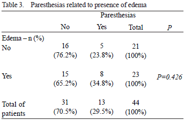
There was no association between edema and paresthesia (value) P=0.4, Chi square test, Table 3.
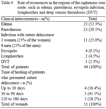
Of the 44 patients, 13 have complained of paresthesia in the operated limb, 11 (eight men and three women) presented infection with suture dehiscence - on which the healing time ranged from 30 to 180 days; four patients presented episodes of erysipelas (two men and two women ), (on which in the two women occurred outbreak and they had family history of lipedema; two patients had lymphorrhea during healing; one patient developed deep vein thrombosis (DVT) in the leg veins 10 days after surgery, and 23 reported appearance of edema in the postoperative period, which was initiated between 30 days and 6 months of surgery (Table 4).
DISCUSSION
This study assessed the main complications in sites of removal of great saphenous vein for coronary bypass. Although there are many studies approaching aspects of CABG, the knowledge of complications that result from the removal of great saphenous vein of the lower limbs is limited. In the traditional technique that is still widely used, the removal of the saphenous vein is performed with a single, long incision along the venous path. The bridged incisions have been performed by reducing the size of incisions in order to reduce complications. Despite the care that aims to simplify the procedure, minimize operative trauma and make the scars cosmetically more attractive in long term, some of these patients have presented severe clinical complications that cause great discomfort in the ipsilateral lower limb.
A study showed an association with paresthesia in 61% of patients [6] assessed and 29.5% of the patients were identified in this study with persistent paresthesia - up to now - that may be related to saphenous nerve lesion during removal of the saphenous vein [11] .
By assessing 1554 patients undergoing coronary artery bypass grafting, 182 patients presented infection in the chest incision and in the lower limb, since the overall rate of postoperative infection - that was 11.7% - of these, 4.6% were superficial infections in the lower limb and 2.2% deep infections in the same region, while in this study were 25% of infection in the saphenous bed. Therefore, this high frequency of infection in the saphenous vein bed in this study provides a warning about such occurrence [12].
The influence of diabetes as a risk factor for the occurrence of complications in the surgical wound was described by many authors [3,11-13] but it was not confirmed in our study. In our study, regarding the appearance of long-term infectious episodes at the operated limb such as lymphangitis and/or erysipelas and its relationship with diabetes, of the 44 patients, four (9.1%) presented episodes of erysipelas: two male (50%) and two females (50%)) on which women were diabetic (50%) and men (50%), were not.
In none of these four patients the presence of tinea
pedis was detected that could have served as entering for bacterial infection. It was mentioned in the study by Greenberg [14], which detected the presence of interdigital mycosis in 100% of the nine patients who presented episodes of lymphangitis.
Only one patient of our sample presented episodes of acute venous thrombosis of deep veins of the leg (fibular veins) - fact occurred 10 days after surgery. As chronic venous insufficiency (CVI) may contribute to the worsening of lymphedema, induce limitation of joint mobility and contribute to edema, patients with prior CVI were excluded from the study [15,16].
Regarding the analysis of presence of edema, it clinically occurred in 23 patients (52.3%), so in more than half of the sample. Of the four patients who presented episodes of erysipelas, all had already developed edema prior to the outbreak, and the two women had family history of lipedema which is a recognized predisposing cause for the onset of lymphedema [17], and in these two women, two episodes of erysipelas occurred in each.
Everything suggests that the presence of edema in these patients should be directly related to the lymphatic rupture, or that is, to the damage suffered during surgery by the parallelism of the important lymphatic channels that accompany the saphenous vein as the ventromedial bundle of the leg and thigh which consistis of the anteromedial or femoral great saphenous vein flow.
CONCLUSION
The bridged excision of the saphenous vein for CABG does not eliminate the early and late clinical complications in the saphenous bed such as paresthesia, infection and edema.
REFERENCES
1. De Milto L, Costello AM. Coronary artery bypass graft surgery. In: Gale encyclopedia of surgery. Gale, Detroit: Gale Group; 2004. Disponível em http://www.healthline.com/galecontent/coronary-artery-bypass-graft-surgery-1. Acesso em: 29/02/2008
2. Favaloro RG. Critical analysis of coronary bypass graft surgery: a 30-year journey. J Am Coll Cardiol. 1998;31(4Suppl B):1B-63B. [MedLine]
3. Tyszka AL, Fucuda LS, Tormena EB, Campos ACL. Obtenção da veia safena magna através de acesso minimamente invasivo para revascularizações miocárdicas. Rev Bras Cir Cardiovasc. 2001;16(2):105-13. View article
4. Bruxton B, Acar C, Suma H. Conduits. In: Bruxton B, Frazier OH, Westaby, editors. Ischemic heart disease surgical management. London: Mosby International; 1999. p.139-77.
5. Reid R, Simcock JW, Chisholm L, Dobbs B, Frizelle FA. Postdischarge clean wound infections: incidence underestimated and risk factors overemphasized. ANZ J Surg. 2002;72(5):339-43. [MedLine]
6. Garland R, Frizelle FA, Dobbs BR, Singh H. A retrospective audit of long-term lower limb complications following leg vein harvesting for coronary artery bypass grafting. Eur J Cardiothorac Surg. 2003;23(6):950-5. [MedLine]
7. Lavee J, Schneiderman J, Yorav S, Shewach-Millet M, Adar R. Complications of saphenous vein harvesting following coronary artery bypass surgery. J Cardiovasc Surg. 1989;30(6):989-91.
8. Mountney J, Wilkinson GA. Saphenous neuralgia after coronary artery bypass grafting. Eur J Cardiothorac Surg. 1999;16(4):440-3. [MedLine]
9. Schoppelrey HP, Breit R. Erysipele nach entnahme von beinvenen für eine aortokoronare bypassoperation. Hautarzt. 1996;47(12):909-12. [MedLine]
10. Dan M, Heller K, Shapira I, Vidne B, Shibolet S. Incidence of erysipelas following venectomy for coronary artery bypass surgery. Infection. 1987;15(2):107-8. [MedLine]
11. Mountney J, Wilkinson GA. Saphenous neuralgia after coronary artery bypass grafting. Eur J Cardiothorac Surg. 1999;16(4):440-3. [MedLine]
12. L'Ecuyer PB, Murphy D, Little JR, Fraser VJ. The epidemiology of chest and leg wound infections following cardiothoracic surgery. Clin Infec Dis. 1996;22(3):424-9.
13. Allen KB, Heimansohn DA, Robison RJ, Schier JJ, Griffith GL, Fitzgerald EB, et al. Risk factors for leg wound complications following endoscopic versus traditional saphenous vein harvesting. Heart Surg Forum. 2000;3(4):325-30. [MedLine]
14. Greenberg J, DeSanctis RW, Mills RM Jr. Vein-donor-leg cellulitis after coronary artery bypass surgery. Ann Intern Med. 1982;97(4):565-6. [MedLine]
15. Cavalheri G Jr, Godoy JM, Belczak CE. Correlation of haemodynamics and ankle mobility with clinical classes of clinical, aetiological, anatomical and pathological classification in venous disease. Phlebology. 2008;23(3):120-4. [MedLine]
16. Godoy JMP, Braile DM, Godoy MFG. Lymph drainage in patients with joint immobility due to chronic ulcerated lesions. Phlebology. 2008;23(1):32-4. [MedLine]
17. Boursier V, Pecking A, Vignes S. Analyse comparative de la Lymphoscintigraphie au cours des lipoedèmes et des lymphoedèmes primitif des membres inferieurs. J Maladies Vasculaires. 2004;29(5):257-61.
Article receive on Sunday, June 1, 2008


 All scientific articles published at rbccv.org.br are licensed under a Creative Commons license